Author
Kim Whitley
Scientific Content Manager
Author
Kim Whitley
Scientific Content Manager
At the International Clinical Cytometry Society (ICCS) Annual Course and Meeting, members of our flow cytometry team, Sanaz Koosha and Nithianandan Selliah, saw these developments reflected across scientific sessions, training courses, and poster presentations. Discussions spanned hematological malignancies, including acute myeloid leukemia (AML), chronic lymphocytic leukemia (CLL), and multiple myeloma (MM), alongside advances in high-parameter assays and automated data analysis.
The two posters from our team explored different aspects of these challenges: one examined comprehensive immunophenotyping panel, a 48-color spectral flow cytometry panel, with AI-assisted analysis across two laboratories, while the other focused on the validation of a flow cytometry panel to characterize AML blast with CD123 quantitation in AML and myelodysplastic syndrome (MDS) patient samples.
Read on for highlights from the event and a closer look at the research our team presented.
One of the clearest themes from ICCS was the continued, growing interest in spectral flow cytometry. More labs are adopting the technology or planning to introduce it, drawn by its ability to measure a broad range of immune cell populations and functional markers in a single tube.
As panels capture more information, making sense of all that data becomes a bigger part of the challenge. Sessions and posters highlighted how artificial intelligence (AI) and machine learning (ML) are being used to support analysis, from automating parts of the workflow to identifying cell populations in complex datasets.
Automation was another key topic, with advances in sample preparation and instrument integration helping reduce manual steps in the lab. Together, AI, ML, and automation continue to shape how flow cytometry data is generated and analyzed. The focus is not just on processing data faster, but on making sure results remain reliable and scientifically meaningful.
Discussions also touched on the use of AI-assisted analysis for measurable residual disease (MRD), where detecting small populations of abnormal cells can be particularly challenging. For these approaches to be used in clinical research, it’s important to show that automated analysis can identify these populations reliably and deliver results that stand up to scientific scrutiny.
High-parameter spectral flow cytometry enables detailed immune profiling, but ensuring data analysis is consistent across labs can be challenging.
Sanaz Koosha presented a poster demonstrating the use of a 48-marker spectral flow cytometry immune profiling panel across two laboratories. The study evaluated cryopreserved PBMC sample and assessed a broad range of immune populations, including B cells, T cells, NK cells, and monocytes. The work combined high-parameter spectral flow cytometry with Ozette Resolve for spectral unmixing and Ozette Endpoints for standardized endpoint analysis.
Following the initial analysis and confirmation of the gating strategy, the workflow can be applied to additional samples, with a second reviewer checking and approving the results. Where adjustments are needed, the analysis can be revisited and refined.
The team tested the workflow at both Cerba Research and Ozette, with different operators and instruments at each site. The results were closely aligned across both laboratories. All four Cerba Research-Ozette run pairings achieved Pearson’s correlation coefficients and Lin’s concordance correlation coefficients greater than 0.99, demonstrating excellent cross-site comparability of the workflow.
Beyond this study, high-parameter spectral flow cytometry panels can be adapted to support different research objectives. Marker selection and panel design can be optimized for specific therapeutic areas or scientific questions, with performance evaluated for the intended application.
The takeaway: More parameters can reveal more about the immune system, but the data needs to be analyzed consistently to make those insights useful. Combining AI-assisted analysis with standardized workflows and expert review can help laboratories work with complex datasets while keeping scientific oversight in place.
Understanding the expression of a target on relevant cell populations can be an important part of patient characterization. This was the focus of Nithianandan Selliah’s poster, which described the validation and implementation of a flow cytometry panel for AML and MDS samples.
The panel focuses on CD123, a target of interest in the associated therapeutic program. Using CD123 quantification with molecules of equivalent soluble fluorochrome (MESF) beads, the assay measures target expression on leukemic blast populations. This provides a quantitative assessment of CD123 expression, helping researchers characterize the cells relevant to the therapy under investigation.
The team evaluated the panel using AML patient samples, assessing key performance parameters including assay precision and sample stability. The results met the predefined acceptance criteria, supporting the assay’s suitability for use in the clinical trial.
The work shows the importance of more than identifying the relevant cell populations. Where a therapy targets a specific receptor, a validated method for quantifying its expression can provide additional information to support the study’s objectives.
The takeaway: Understanding how much of a target such as CD123 is expressed on cancer cells can help researchers assess its relevance to targeted therapies. Validated assays and reliable reference materials help make those measurements consistent and meaningful.
ICCS showed how spectral flow cytometry, AI-assisted analysis, and automation are changing the way laboratories study immune cells. But getting more information from each sample is only part of the picture. The results also need to be consistent, and assays need to be validated for their intended use.
The two posters from our team show different aspects of this challenge:
Both underline the importance of combining advanced technology with rigorous scientific methods. As these approaches become more widely used, the priority is making sure they deliver data researchers can rely on to support clinical research.
Explore our flow cytometry capabilities or contact us to discuss your clinical research requirements.
Cerba Research and Ozette Technologies Announce Partnership to Advance High-Dimensional Immune Profiling and Improve Clinical Decision-Making

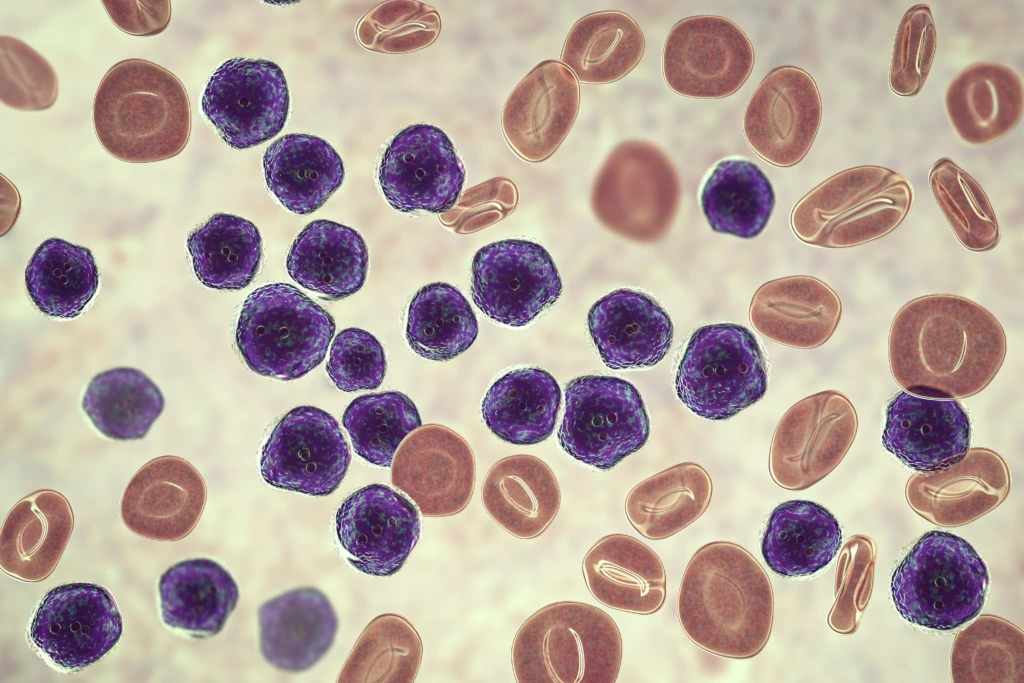
Learn how multiparameter flow cytometry supports AML clinical trials, from patient selection and receptor occupancy to measurable residual disease (MRD) monitoring.
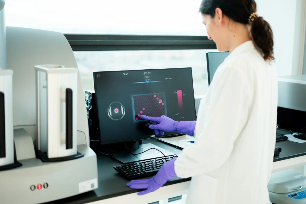
In this interview, NewsMedical talks to Cerba Research’s Coen Stalpers about advances in automation and artificial intelligence in the laboratory setting.
Learn how advanced flow cytometry assays support key activities such as CAR-T cell tracking, biomarker identification, treatment response assessment, and minimal residual disease (MRD) evaluation, helping to generate high-quality data and drive informed decision-making in complex cell therapy trials.