
Author
Sanaz Koosha
Scientist, Flow Cytometry
Author
Sanaz Koosha
Scientist, Flow Cytometry

Author
Rowan Claeys
Clinical Pathologist
Acute myeloid leukemia (AML) is a genetically and biologically heterogeneous hematologic malignancy. Recent WHO and ICC classifications recognize recurrent genetic abnormalities as a cornerstone of AML diagnosis, often carrying greater diagnostic and clinical significance than the traditional blast cell threshold (Platzbecker et al., 2025; Döhner et al., 2022). These genetic alterations not only determine risk stratification and prognosis but increasingly guide targeted therapies and, for selected subtypes, enable molecular monitoring of measurable residual disease (MRD). As AML treatment becomes more personalized, clinical trials require equally precise tools to evaluate treatment response at the cellular level.
Genomic sequencing provides a comprehensive view of a patient’s genetic landscape but cannot capture cellular behavior in real time. Multiparameter flow cytometry (MFC) serves as the “eyes on the ground.” It tracks cellular behavior (phenotype), verifies drug binding to blast cells (receptor occupancy (RO) assay), and measures the true depth of remission (by MRD assay). From first enrollment to long-term follow-up, flow cytometry acts as the ultimate compass in modern AML drug development (Jum’ah et al., 2025).
Historically, AML trials used a simple visual threshold for remission. Doctors looked under a microscope to see if leukemic blasts dropped below 5%. Today, we know this visual check is far too blunt. A patient can pass this test and still harbor millions of cancer cells. These residual leukemic cells, including therapy-resistant leukemic stem cells capable of reinitiating disease, are the primary drivers of relapse (Schuurhuis et al., 2018).
While intensive chemotherapy remains the backbone of AML treatment, the pipeline of new drugs has exploded with specialized options:
With such targeted treatments, researchers need deeper data. They must confirm a target exists before a patient enrolls. They need to see if the drug actually binds to that target, and they must measure cancer elimination far beyond what a microscope can see. Flow cytometry provides this information rapidly and reliably (Jum’ah et al., 2025).
An AML trial depends on matching targeted therapies to the patient’s disease biology. Flow cytometry screens bone marrow and blood samples to identify the appropriate immunophenotypic targets (Jum’ah et al., 2025).
Finding leukemic blasts
With flow cytometry, combining forward scatter (cell size), side scatter (internal complexity), and multiparameter fluorescence, AML blasts can be distinguished from normal hematopoietic populations based on their characteristic immunophenotypic profile (Jum’ah et al., 2025).
Mapping cellular blueprints
Once identified, AML blasts are characterized by their immunophenotypic profile, revealing key abnormalities common in AML (Jum’ah et al., 2025):
This detailed map supports AML diagnosis and identifies potential therapeutic targets for patient selection, helping to avoid unnecessary side effects, and provides the phenotypic fingerprint used for subsequent MRD monitoring.
Before treatment begins, researchers build a customized tracking plan for each patient (Schuurhuis et al., 2018).
Creating cellular fingerprints
The cornerstone of this step is identifying leukemia-associated immunophenotypes (LAIPs). These are unique, abnormal combinations of markers found on the patient’s cancer cells that create an individual phenotypic fingerprint (Schuurhuis et al., 2018).
Documenting this baseline immunophenotype before treatment is essential. As healthy bone marrow cells regrow after therapy, researchers must know exactly what the original cancer looked like to spot any residual leukemic cells.
Measuring disease burden
Flow cytometry quantifies the leukemic blast burden at diagnosis, establishing a baseline against which treatment response and disease clearance can be monitored throughout therapy (Mawalankar et al., 2025; Jum’ah et al., 2025).
Modern AML treatments rely heavily on flow cytometry to judge success, watch recovery, and track how different drugs function (Platzbecker et al., 2025). Complementing molecular techniques, flow cytometry provides MRD assessment for most AML patients, including those without a suitable genetic marker for molecular follow-up .
Intensive chemotherapy
During high-dose chemotherapy, flow cytometry helps monitor bone marrow recovery by distinguishing regenerating hematopoiesis from persistent residual leukemia, helping clinicians differentiate treatment-related aplasia from active marrow regeneration (Kern et al., 2010).
Low-intensity combinations
For patients who cannot tolerate intensive chemotherapy, doctors often pair venetoclax, a BCL-2 inhibitor, that promotes apoptosis with other agents. Flow cytometry acts as a real-time tracker, monitoring treatment response and changes in cellular populations throughout therapy (Platzbecker et al., 2025).
Smart targeted therapies
For specific genetic mutations, clinicians use targeted inhibitors (Döhner et al., 2022):
In these trials, flow cytometry assays measure exactly how well the drug binds to and blocks its targets (receptor occupancy assay) and monitors blast populations (Jum’ah et al., 2025).
Stem cell transplants
The success of a stem cell transplant depends entirely on achieving a deep remission beforehand. If flow cytometry detects lingering cancer fingerprints before the procedure, the risk of relapse rises significantly (Cloos et al., 2026; Demir et al., 2026). This alerts doctors to adjust pre-transplant therapy or introduce fallback treatments.
AML creates a hostile environment in the bone marrow that paralyzes healthy immunity. When a trial introduces an immunotherapy, MFC is used to evaluate the entire immune battlefield (Demir et al., 2026).
Researchers can analyze multiple immune populations in a single sample (Demir et al., 2026):
Flow cytometry also checks activation markers to see if immune cells are actively fighting while tracking checkpoint receptors to flag immune exhaustion and guide combination drug strategies (Demir et al., 2026).
Measurable Residual Disease (MRD) refers to the tiny number of cancer cells that remain after a patient achieves a standard remission. Reaching an “MRD-negative” state is one of the strongest predictors of relapse and long-term survival in AML (Schuurhuis et al., 2018). Because of this, MRD status is increasingly used as a surrogate endpoint to accelerate new drug approvals.
Trials use two primary flow-based methods to spot MRD (Schuurhuis et al., 2018; Cloos et al., 2026; Jum’ah et al., 2025):
A standard microscope can only detect cancer down to about 5% (1 in 20 cells). Flow cytometry routinely achieves a sensitivity of 10-4 to 10-5(1 in 10,000 to 1 in 100,000 cells). This provides the depth to confidently find a single remaining leukemia cell hidden among a large number of healthy cells (Schuurhuis et al., 2018; Cloos et al., 2026).
According to the updated ELN recommendations, MFC-MRD positivity is defined as ≥0.1%, while lower levels of detectable disease above the assay’s limit of quantification (MRD-low level) are increasingly recognized as potentially clinically relevant and are the subject of ongoing investigation (Cloos et al., 2026).
Standardized flow cytometry is essential to generate reliable and comparable MRD results across clinical trial sites. The 2025 ELN-DAVID recommendations provide guidance on validated MFC-MRD assays, harmonized implementation of the integrated LAIP/DfN approach, standardized reporting, and participation in external quality assessment (EQA) programs to ensure robust and clinically meaningful MRD assessment (Cloos et al., 2026). Complementary ELN-DAVID technical recommendations address standardized antibody panels, instrument harmonization, data acquisition, gating and data analysis, and quality control (Wood, van der Velden, Buccisano et al.). These efforts are further supported by initiatives such as the EuroFlow Consortium, which has pioneered standardized protocols and data analysis strategies to improve reproducibility across laboratories (Cloos et al., 2026).
Future precision medicine approaches will increasingly integrate flow cytometry with genomic, transcriptomic, and proteomic data, providing a more comprehensive understanding of disease biology and treatment response.
Spectral flow cytometry
Conventional flow cytometry already enables high-parameter immunophenotyping, while spectral flow cytometry further expands panel design by capturing the full emission spectrum of each fluorochrome. This allows researchers to simultaneously measure 40+ markers, enabling comprehensive mapping of leukemic cells, healthy hematopoietic populations, and the immune system within a single sample (Spasic et al., 2024).
AI/machine learning-assisted data analysis
Integrating machine learning algorithms is revolutionizing data analysis workflows (Zhang et al., 2025). AI tools can analyze complex data shapes simultaneously to group cell populations automatically. This increases reporting speed, eliminates human bias, and spots ultra-rare leukemia stem cells that human experts might miss.
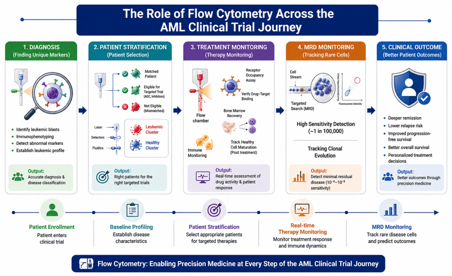
MFC ensures AML trials enroll the right patients, verifies drug targets, maps immune responses, and delivers high-sensitivity data to prove clinical success. Through standardized testing, spectral advancements, and machine learning, flow cytometry remains an indispensable pillar for developing safer, more effective targeted cures.
References
We’re exploring one of the most important topics in flow cytometry: building quality into your flow cytometry data. At first glance, this may seem like a straightforward topic. However, there are many sources of variability that can find their way into your data and ultimately affect the consistency of your results.

Read our key takeaways from ISCT 2026, from in vivo CAR-T and autoimmune applications to evolving regulation in cell and gene therapy.

Learn more about key oncology trends from AACR 2026 including AI-driven pathology, liquid biopsy advances, antibody therapeutics, and immune profiling in cancer research.
Explore advances in spectral flow cytometry, AI-assisted data analysis, immune profiling, and validated flow cytometry assays for clinical research.