Get in touch with us
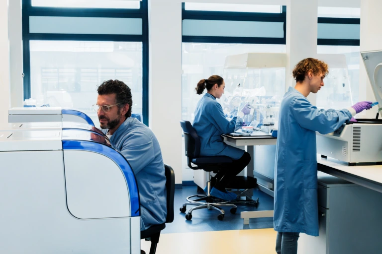
Scientists working together in laboratory wide shot

Driving Translational Research with Deeper Tissue Insights

Drive confident decisions with integrated histopathology, advanced imaging, and expert-led tissue analysis across global clinical programs.

Consistency across complex tissue-based studies

Modern clinical research demands more than traditional pathology. Today’s programs rely on deep tissue insights, advanced biomarker characterization, and reproducible data that can scale across global trials.

To generate decision-ready evidence, you need harmonized workflows, validated assays, and scientific expertise that connects tissue-level observations to clinical outcomes.

We provide end-to-end anatomic pathology and tissue analysis solutions, supporting your program from early assay development through to validated clinical trial testing across therapeutic areas like oncology, neurology, and beyond.

Our integrated capabilities include:

  • Immunohistochemistry (IHC) and immunofluorescence (IF)
  • Multiplex histology and spatial biology
  • Digital pathology and image analysis
  • Tissue processing, characterization, and biobanking
  • Pathologist-led interpretation and reporting.

With globally harmonized laboratories and standardized processes and platforms, we deliver consistent, high-quality data you can rely on at every stage.

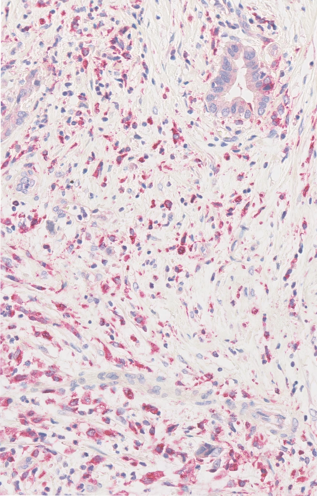
CD68 (Clone KP1) IHC staining of Non-Small Cell Lung Cancer FFPE sample.

Supporting your research from discovery to clinical translation

Our integrated anatomic pathology, imaging, and tissue analysis capabilities help generate meaningful insights across the drug development continuum, supporting:

  • Biomarker discovery and validation to identify and characterize clinically relevant tissue markers 
  • Mechanism-of-action studies to understand biological responses to therapeutic intervention
  • Translational research and endpoint analysis to connect tissue-level findings with clinical outcomes
  • Patient stratification strategies to identify responsive populations and support precision medicine approaches 
  • Companion diagnostic development through robust assay development and validation
  • Longitudinal tissue-based analysis to monitor biological changes over time and throughout treatment
M2 Headers (1)

Delivering the expertise your program demands

Expert-led scientific partnership

Scientist leading collaborative laboratory discussion
  • IHC and IF assay development, optimization, and validation
  • Guidance on biomarker strategy and study design
  • Access to experienced pathology experts 
  • Expert support from assay development through clinical trials 
  • Integrated histopathology, imaging, and biomarker expertise

Advanced tissue and spatial analysis

shutterstock_1255890574-1-scaled.jpg
  • Multiplex histology and imaging approaches
  • Spatial characterization of complex biological systems
  • High-quality image analysis using advanced software

Scalable global delivery

Female warehouse worker checking stock inventory A
  • Harmonized SOPs across global laboratory network
  • Standardized workflows and quality control processes
  • Proven delivery across multi-site clinical studies 
  • Reproducible, high-quality data at scale
  • Global regulatory compliance and audit readiness

Tailored to your study needs

Portrait of male scientist during laboratory discussion
  • Custom and off-the-shelf biomarker assays
  • Fit-for-purpose validation strategies
  • Flexible assay development and technology transfer

Experience you can rely on

60+
labs network across the globe
Duotone_Globe
250+
validated tissue-based biomarkers available
Duotone_Assay-Development
25+
sourcing partners for sample access
Duotone_Hand-and-Cog

Comprehensive anatomic pathology and tissue analysis capabilities

IHC and IF

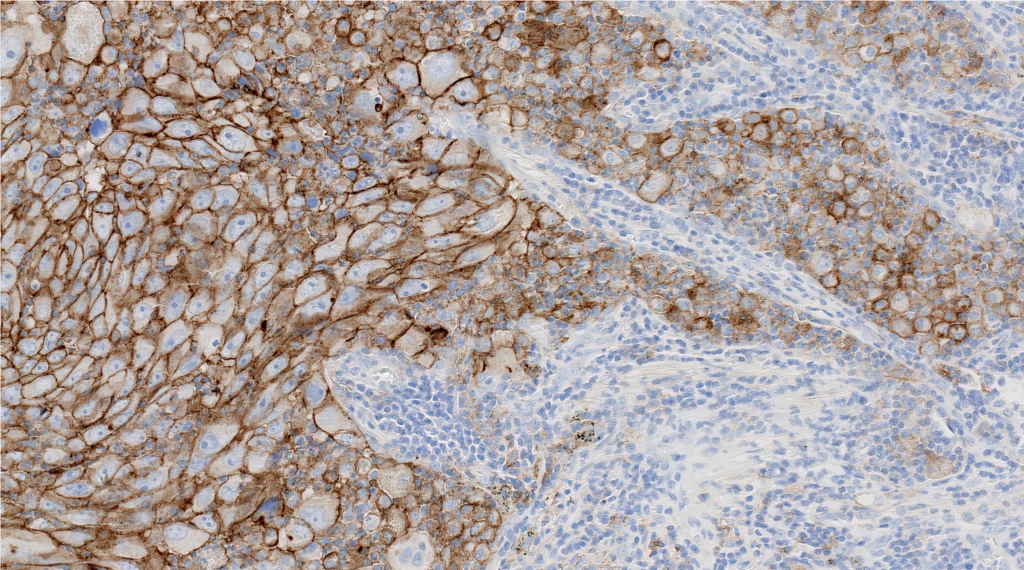
PD-L1 IHC 22C3 pharmDx (Clone 22C3) IHC staining of Non-Small Cell Lung Cancer FFPE sample.

Develop robust, reproducible assays using validated methods across global laboratories, ensuring consistency from early development to clinical application. Access our catalog of 250+ validated assays and custom development solutions.

Histopathology and tissue profiling

Frame_29_2x-2

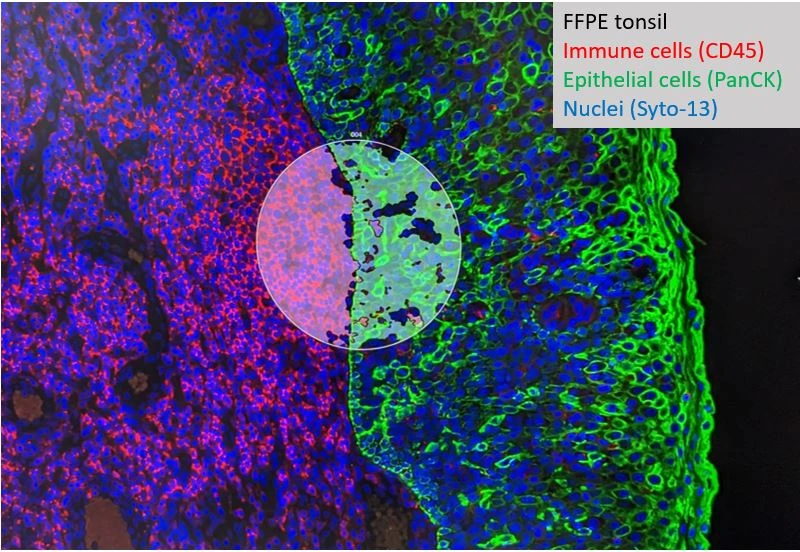
Generate deeper insight from tissue samples through multiplex staining and advanced imaging, enabling simultaneous analysis of multiple biomarkers within a single sample. Stain multiple proteins at once for complex phenotyping and visualization of cell-cell interactions.

Digital pathology and image analysis

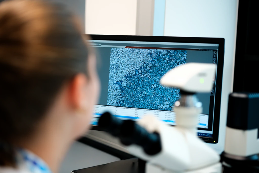
Scientist reviewing microscope imaging data on monitor

Leverage digital pathology platforms and AI-supported image analysis tools to quantify biomarker expression and tissue architecture with precision and reproducibility. Find out about our solutions for advanced image analysis and for digital pathology.

Antibody selection and validation (Histoselect®)

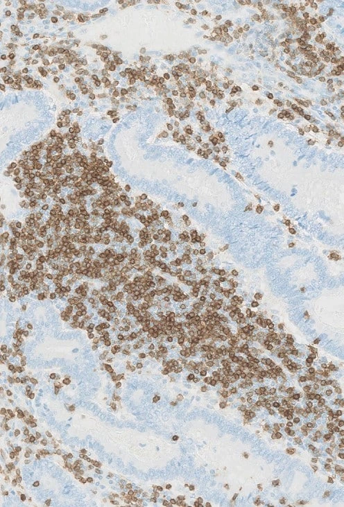
CD3-IHC-staining-of-colorectal

Accelerate antibody development with high-throughput screening capabilities:

  • Screen over 1000 antibody clones per day
  • Evaluate multiple clones and controls on a single slide
  • Identify optimal IHC-performing antibodies efficiently

Tissue processing and biobanking

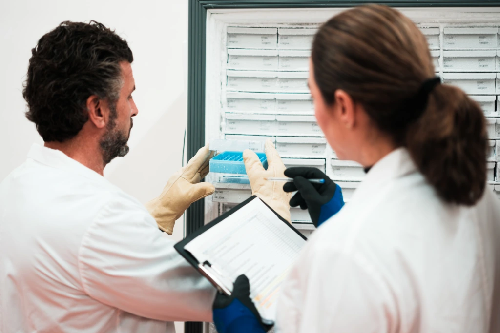
Two scientists reviewing cold storage samples A

Access high-quality biological samples, including FFPE tissues and control materials, supported by standardized processing and storage workflows to ensure data integrity.

In situ hybridization (ISH) assay and RNAscope®

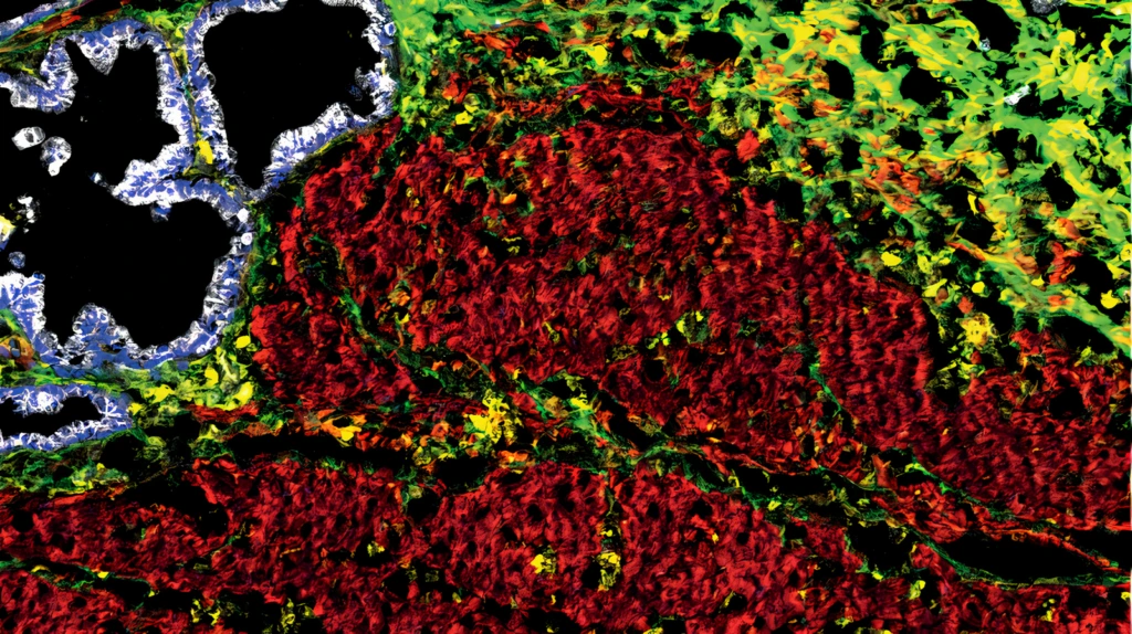
Representative image of 4 targets in a 12plex. Alpha-SMA (Clone 1A4, Red), Vimentin (Clone RV202, Yellow), PanCK (Clone C11, White), Collagen I (Polyclonal, Lime green) multiplex IMC staining of Frozen Pancreas sample. Platform: Discovery ULTRA.

Detect RNA in tissue sections through both simplex and multiplex solutions.

Spatial omics

OMICS

Bring your studies to the next level with spatial transcriptomics and proteomics.

Early phase and preclinical tissue solutions

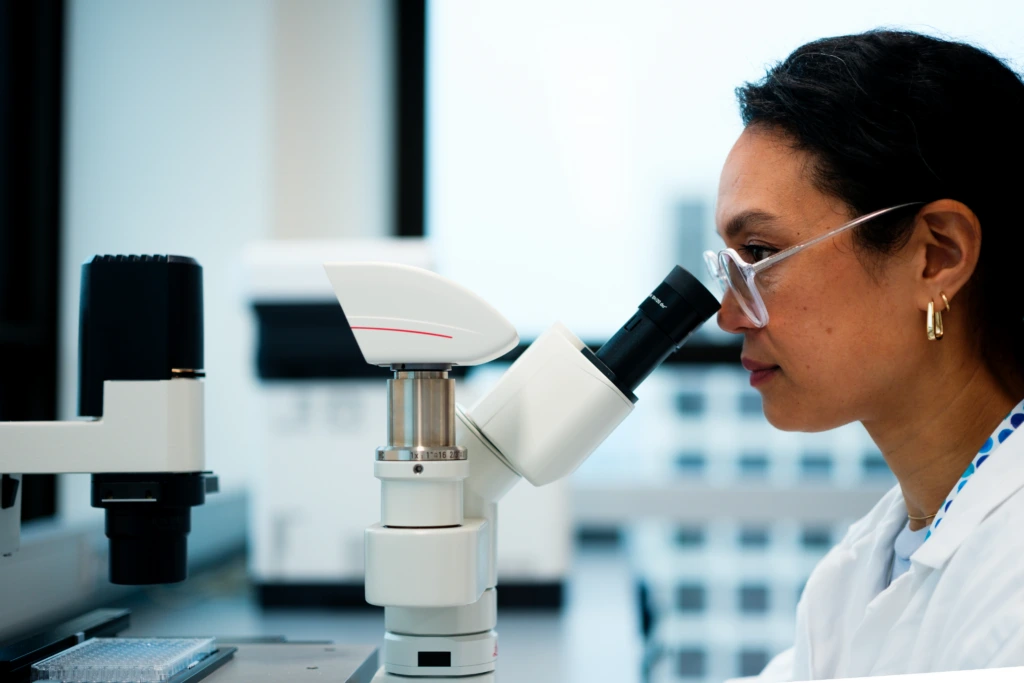
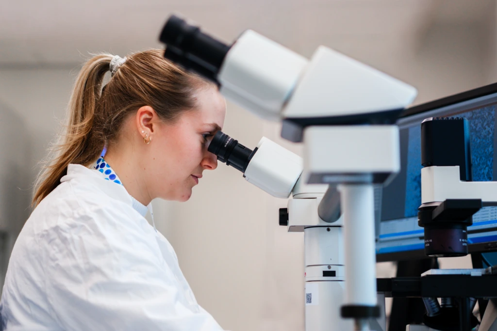
Scientist performing microscopy analysis in laboratory

Discover our offers for histopathology on animal models and tissue cross-reactivity analysis.

Ready to strengthen your tissue analysis strategy?

Connect with our scientific experts to design and deliver robust anatomic pathology and tissue analysis solutions tailored to your program needs.

Resources and insights for anatomic pathology and tissue analysis

Poster

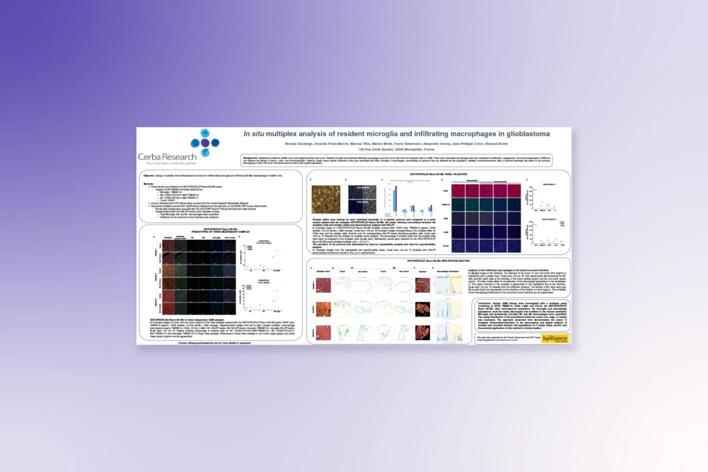
In Situ Multiplex Analysis of Microglia and Macrophages in Glioblastoma

Explore how multiplex spatial analysis differentiates resident microglia and infiltrating macrophages within the glioblastoma tumor microenvironment.

In Situ Multiplex Analysis of Microglia and Macrophages in Glioblastoma
Poster

Multiplex Immunofluorescence Detection of Resident Memory T Cells in Solid Tumors

See how advanced multiplex IF enables precise detection and spatial mapping of resident memory T cells across solid tumor tissues.

Multiplex Immunofluorescence Detection of Resident Memory T Cells in Solid Tumors
Poster

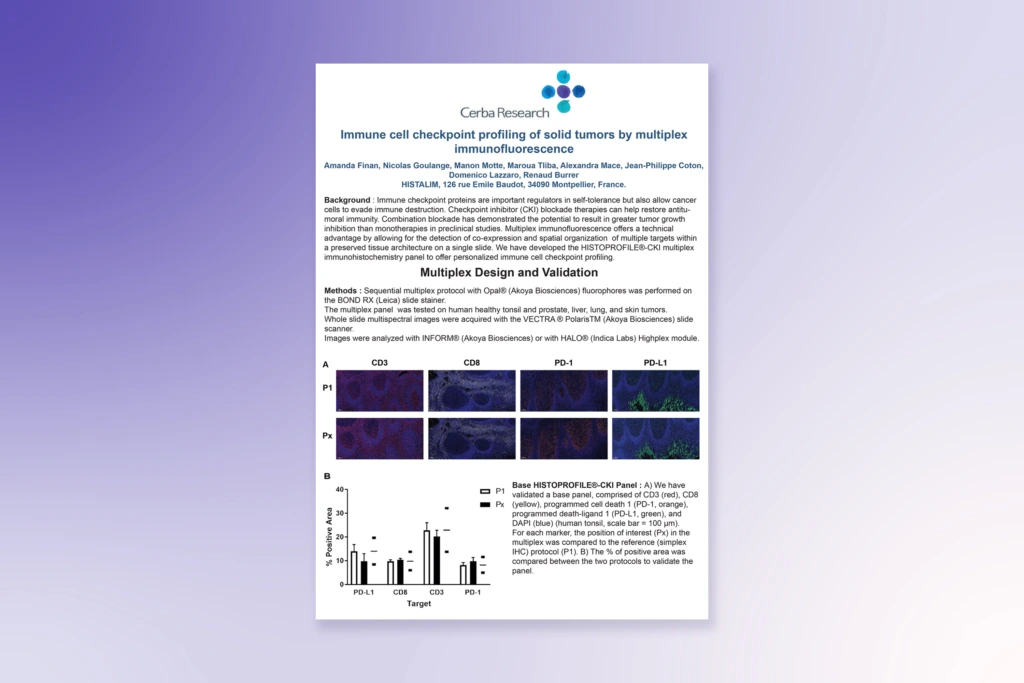
Immune Cell Checkpoint Profiling by Multiplex Immunofluorescence

Understand how multiplex IF supports detailed immune checkpoint profiling within complex tumor microenvironments.

Immune Cell Checkpoint Profiling by Multiplex Immunofluorescence
Poster

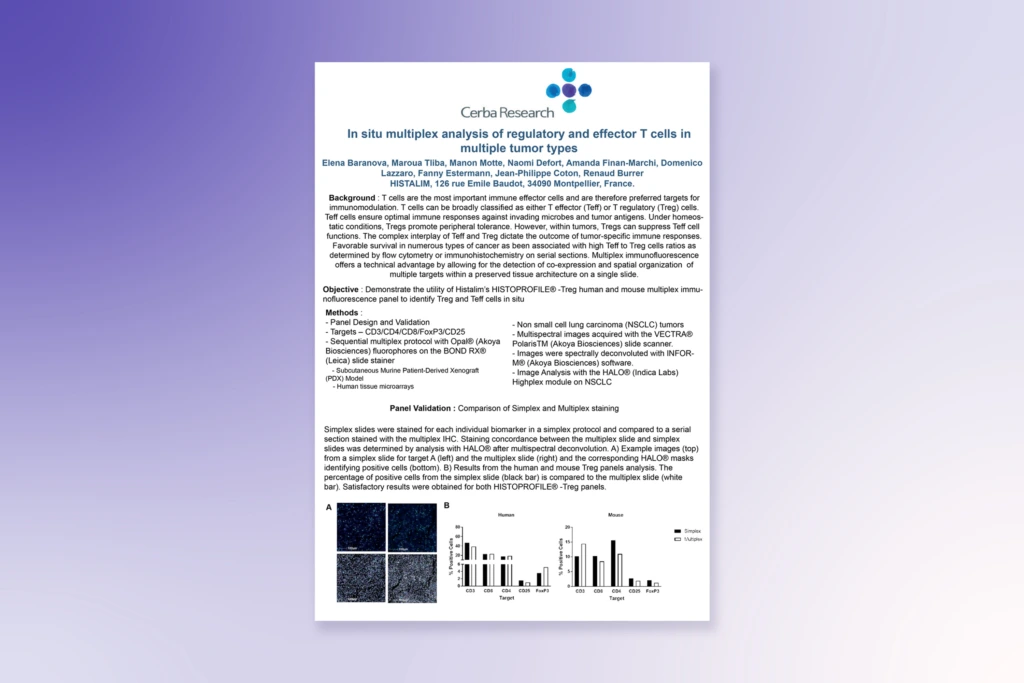
In Situ Multiplex Analysis of T Cells Across Multiple Tumor Types

Discover how spatially resolved multiplex analysis reveals regulatory and effector T-cell interactions within the tumor microenvironment across multiple tissue types.

In Situ Multiplex Analysis of T Cells Across Multiple Tumor Types
Poster

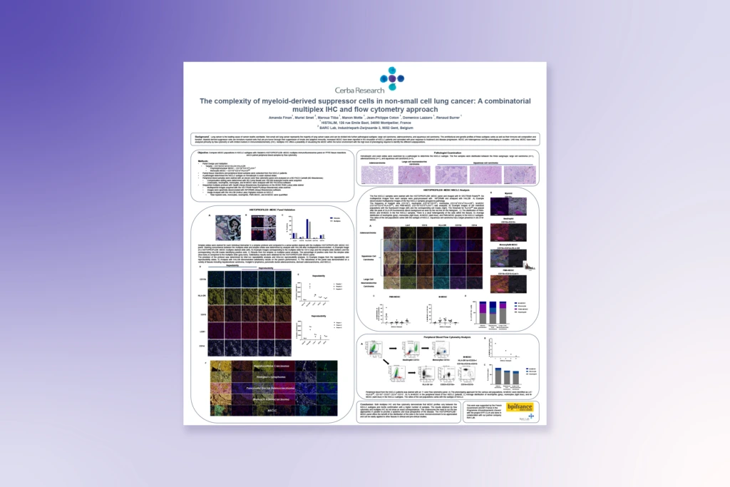
Multiplex IHC Characterization of Myeloid-Derived Suppressor Cells in NSCLC

Learn how a combined multiplex IHC and flow cytometry approach enables comprehensive profiling of immunosuppressive cells in lung cancer.

Multiplex IHC Characterization of Myeloid-Derived Suppressor Cells in NSCLC
Poster

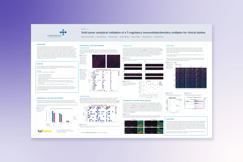
Analytical Validation of a T-Reg Multiplex IHC Panel for Solid Tumors

See how validated multiplex IHC panels support reproducible identification of regulatory T cells in clinical oncology studies.

Analytical Validation of a T-Reg Multiplex IHC Panel for Solid Tumors