End-to-end oncology expertise

- Extensive experience across solid tumors, from first-in-human to phase IV
- Specialized support across tissue, cell, DNA/RNA, and protein modalities
- Contributions to the approval and expansion of oncology therapies
Deliver the biomarker data and integrated insights needed to guide decisions, reduce delays, and keep your oncology program moving toward patients.
Early‑phase trials now depend on complex, multimodal biomarkers, while timelines continue to compress. What’s needed is a scientifically integrated approach that brings together tissue, cellular, molecular, and spatial data into a single, coherent view of tumor biology.
Our scientific teams work with you to design and deliver that approach, aligning biomarker strategy, assay selection, and global laboratory execution to your study objectives and endpoints, without compromising data quality or turnaround times.
We support solid tumor programs through a fully integrated oncology toolkit, including histopathology and immunohistochemistry (IHC), next-generation sequencing (NGS), digital pathology, high-parameter flow cytometry enhanced with AI-driven analytics, and spatial biology.
With experience across more than 360 oncology trials, we deliver the data you need when you need it – so you can focus on turning insights into therapies that truly impact patients’ lives.
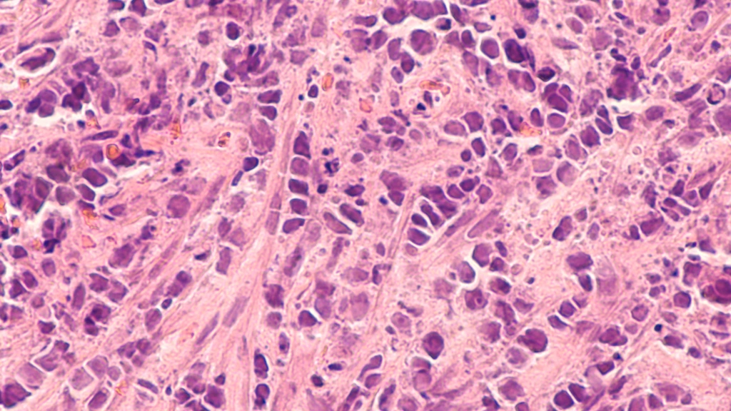
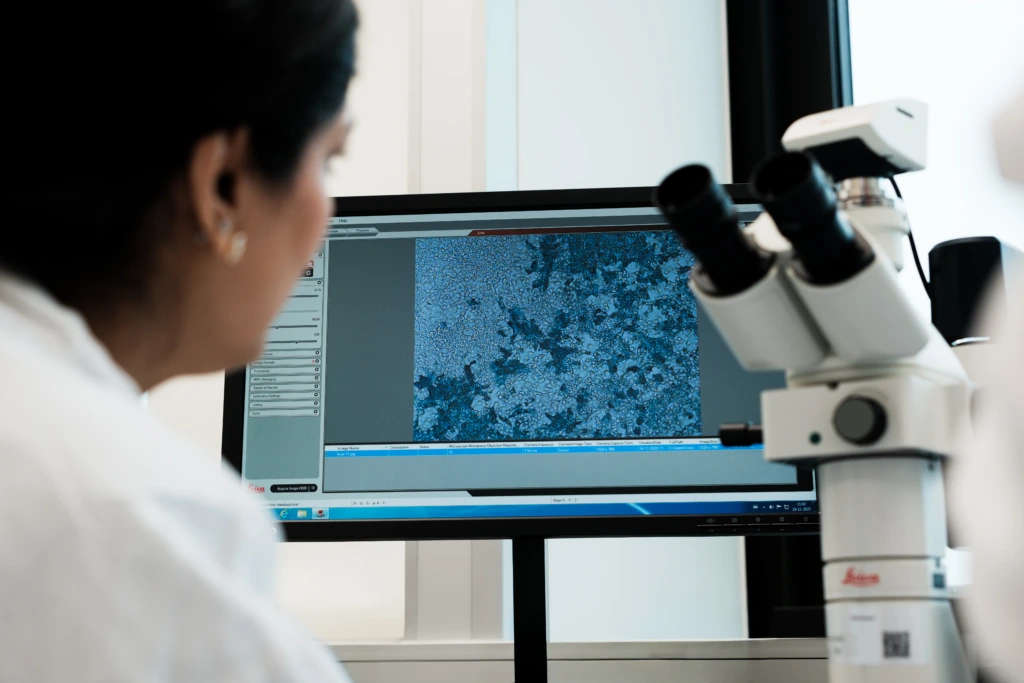


By integrating genomic, phenotypic, proteomic, and morphological insights, supported by advanced computational analytics, we help you turn complex data into clear, confident decisions at every stage of your program.
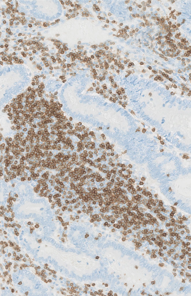
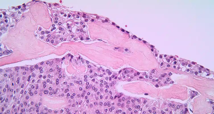
The tumor microenvironment shapes how patients respond, and sometimes resist, therapy. Our global histopathology network delivers high-quality tissue processing, expert morphology review, and advanced singleplex and multiplex IHC panels tailored to solid tumors, helping you turn complex tissue data into actionable insights.
Techniques and approaches include:
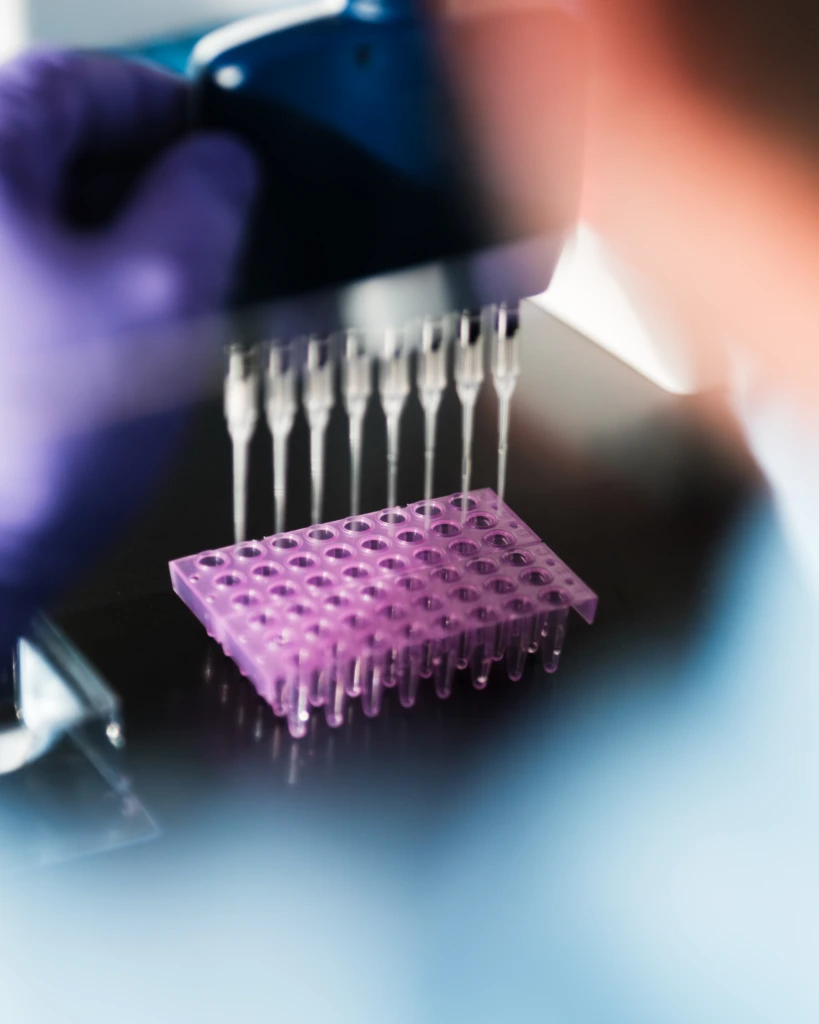
Understanding the genetic and molecular drivers of tumors is key to precision medicine. Our molecular oncology teams deliver high-fidelity sequencing, mutation profiling, and transcriptomic analysis that translate complex genomic data into actionable insights for your trials.
Techniques and approaches include:
Solid tumor trials require deep, multidimensional immune profiling to capture therapeutic effects and patient variability. Our immunology and flow cytometry capabilities illuminate immune activation, suppression, and cellular responses, giving you a complete view of how therapies interact with the immune system.
To further enhance high-dimensional immune profiling, we integrate advanced spectral flow cytometry with AI-driven analytics. This enables standardized, high-parameter immune profiling combined with cloud-based data analysis, helping you translate complex single-cell data into clear, reproducible insights across studies and sites.
Techniques and approaches include:
Accurate measurement of soluble biomarkers, PK, and immunogenicity is critical for biologics, cell therapies, and immunotherapies. Our harmonized global workflows give you reliable, high-quality data while reducing operational burden, so your team can focus on translating results into patient impact.
Techniques and approaches include:
Specialized immunogenicity assessments, including vector-specific and cell and gene therapy biomarker workflows






Our globally harmonized labs and Centers of Excellence deliver reliable, decision-ready data for solid tumor programs worldwide.
Histopathology/IHC:
United States, France, Taiwan, China
Genetics and genomics:
France, Netherlands, Taiwan, United States, Canada
We use state-of-the-art technologies to generate integrated, high-resolution data that drives confident clinical decisions for your program.
Conventional flow cytometry
High‑parameter flow cytometry
AI-driven analytics for flow cytometry
Spatial biology and digital spatial profiling
Whole-slide imaging
Digital pathology and image analysis
Next-generation sequencing (NGS)
Every solid tumor program needs fit-for-purpose and analytically reliable biomarkers. To support this, we maintain a broad menu of validated and exploratory IHC biomarkers.
The following lists summarize the simplex and multiplex markers most frequently used in our oncology trials, with customization available when required.
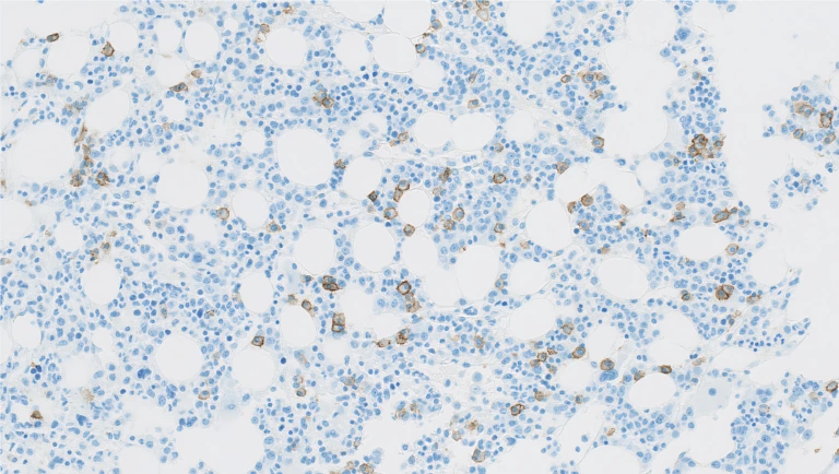
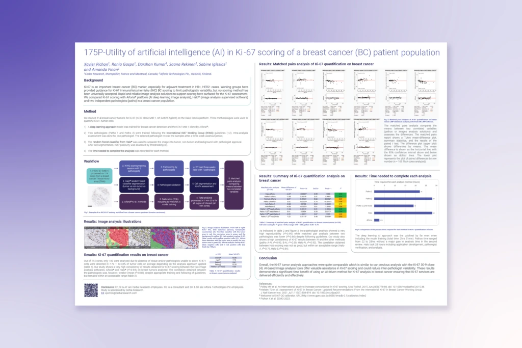
This poster presents data from our study evaluating AI-driven image analysis for Ki-67 quantification in a cohort of 114 breast cancer tumors.
Using the Aiforia® deep learning platform, Halo® image analysis, and two independently trained pathologists, we compared speed, reproducibility, and consistency across scoring methods. The findings show that AI delivers highly consistent Ki-67 results, matching both manual and software-based approaches while dramatically reducing analysis time.
This demonstrates the potential of AI to support more efficient, reproducible Ki-67 assessments, which is especially valuable in HR+/HER2- breast cancer cases where accurate proliferation scoring informs treatment decisions.
Get a clear, data‑driven snapshot of our solid tumor capabilities in this fact sheet summarizing the breadth of our oncology expertise. Explore our comprehensive assay portfolio and learn how our integrated oncology capabilities can support your next trial.

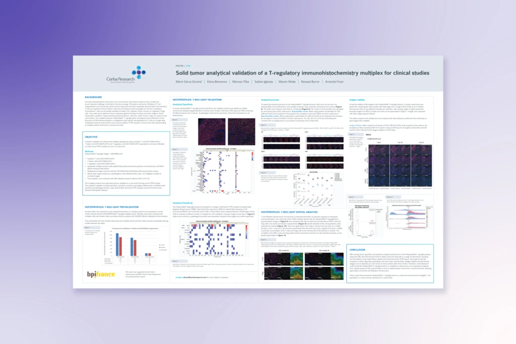
This poster showcases our analytical validation of the HISTOPROFILE®-T-reg light multiplex IHC panel, a fluorescent, multi-marker assay designed to quantify various T cells within solid tumor FFPE tissue. Explore how this validated multiplex can elevate your immune-profiling strategy.
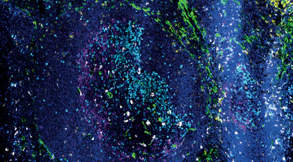
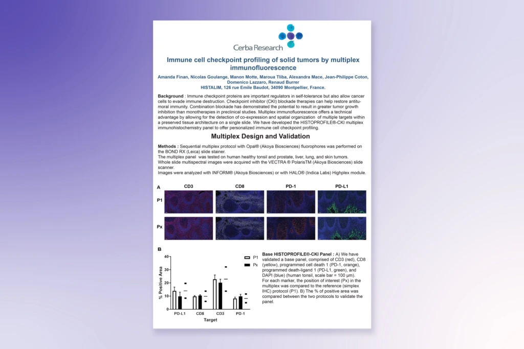
How is multiplex immunofluorescence (IF) reshaping immune checkpoint profiling in solid tumors? In this poster, we present the development and validation of a multiplex assay designed to measure key checkpoint markers and visualize their spatial distribution within intact tumor architecture.
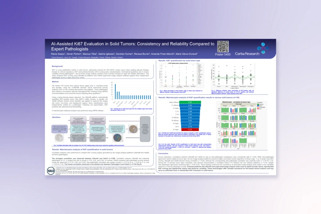
See how AI-assisted image analysis improves reproducibility and quantitative accuracy of Ki-67 scoring across solid tumor tissue samples.
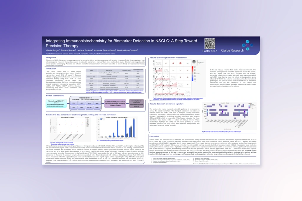
Explore how IHC-based screening of actionable targets supports treatment selection in NSCLC, with reflex testing to improve diagnostic accuracy where needed.
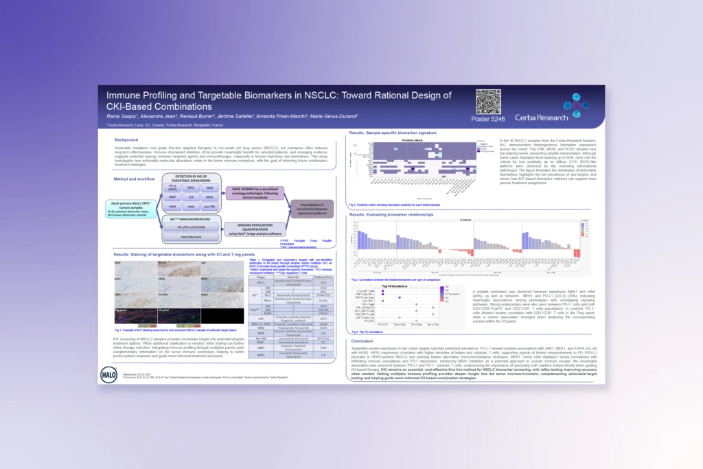
Find out how combining IHC biomarker screening with multiplex immune profiling reveals tumor-immune relationships that can guide more informed checkpoint inhibitor combination strategies.
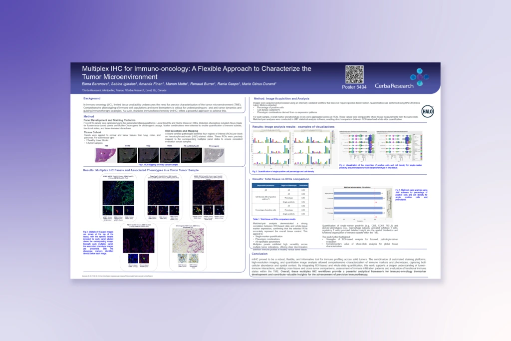
See how five multiplex IHC panels were developed and validated to profile immune cell populations, functional states, and tumor-immune interactions across solid tumor types.