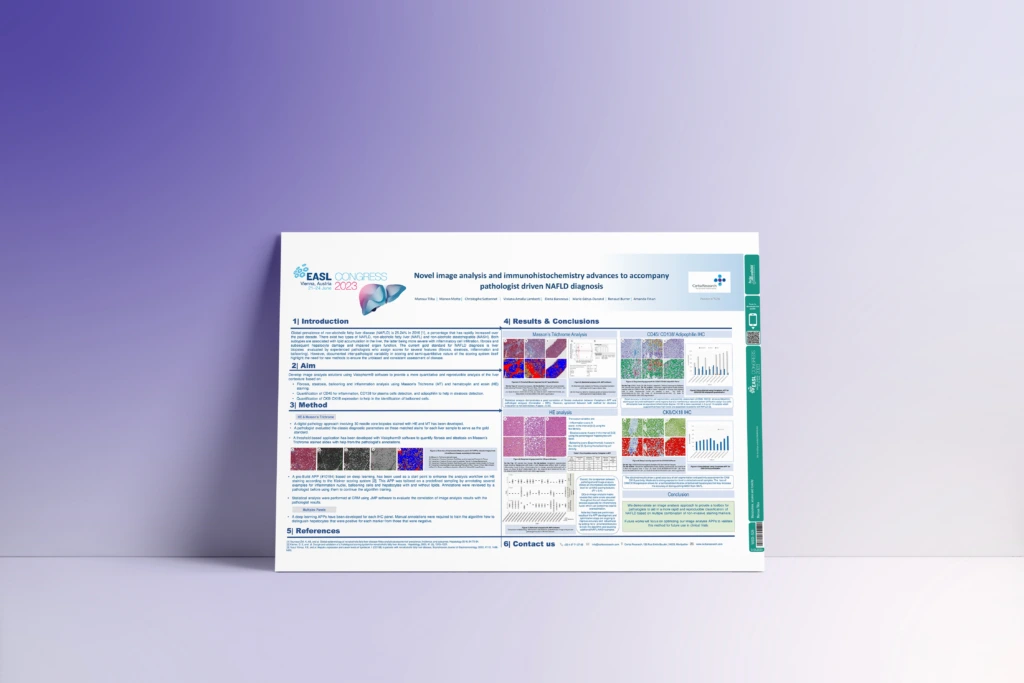
AI-Driven Ki-67 Analysis in Solid Tumors
Discover how AI-powered image analysis enhances the accuracy and reproducibility of Ki-67 scoring for tumor proliferation assessment.
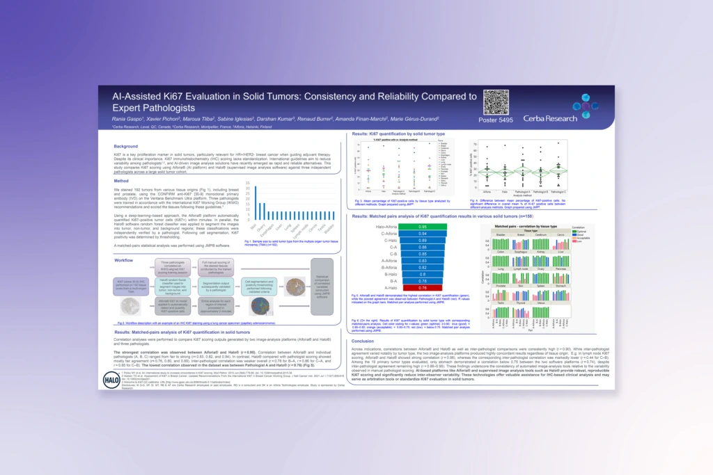
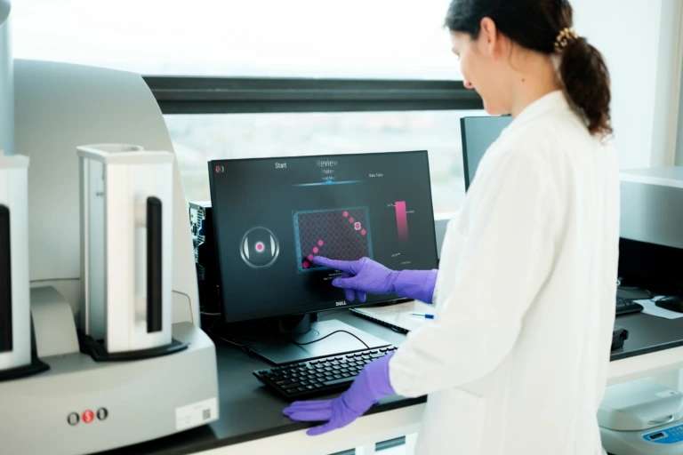
Unlock deeper insights from tissue data with advanced digital pathology and image analysis.
A scanned slide is just the start. To get data you can trust, you need validated platforms, consistent workflows, and pathologists who can identify meaningful biological findings from complex tissue images.
From digitization to AI-powered quantification and spatial analysis, we help you generate reproducible, site-to-site consistent data that helps answer critical questions in your trial.
Our global lab network brings together high-throughput imaging, remote review, and multiplex capabilities to support your biomarker-driven studies across therapeutic areas, generating robust image data that can stand up to rigorous scientific and regulatory scrutiny.

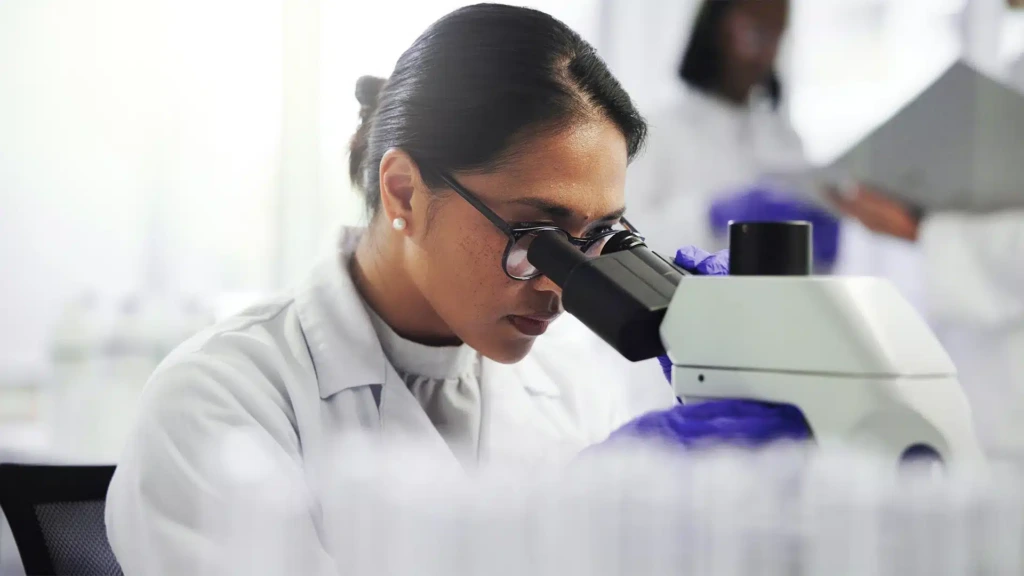
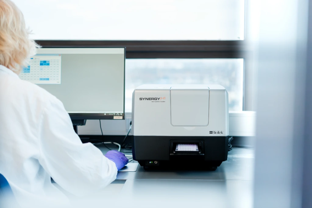
All our tissue analysis laboratories are equipped with brightfield (BF) scanners that are validated for use at a clinical grade. Multispectral digitization is available both in the United States and Europe. The PhenoImager© from Akoya allows fast scanning of samples stained with up to eight different biomarkers in immunofluorescence.
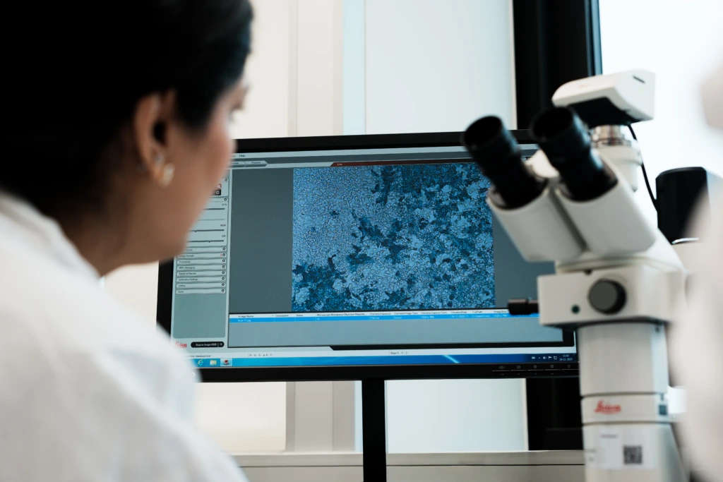
Getting the best data from stained histological sections cannot always rely on pathologist readings, particularly when multiplexing solutions are required. Our image analysis team has been dedicated to providing custom and flexible solutions to extract quantifiable data from biopsy images since 2014. They will suggest expert approaches to get the best dataset from your samples.
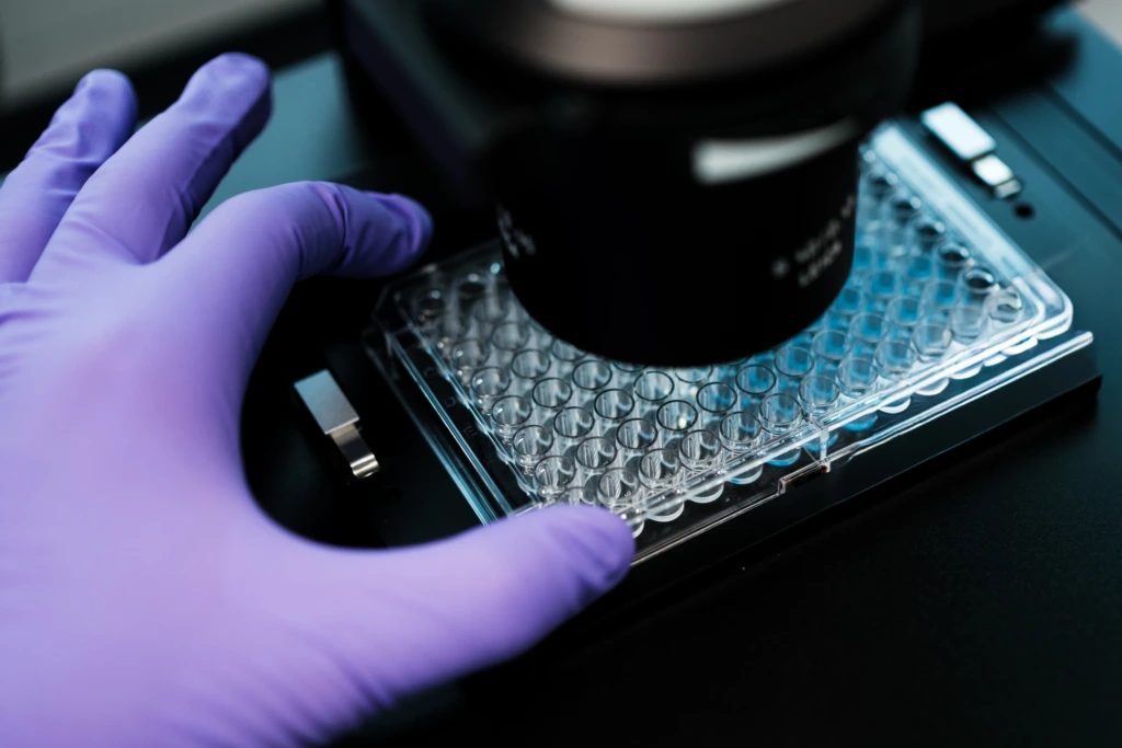
Our multiplexing capabilities allow for accurate phenotyping of cell subpopulations based on the expression or absence of expression of combinations of biomarkers, just as with flow cytometry.
Moreover, additional data on the spatial organization of the tissue can be obtained, providing information on cell interactions that are critical, especially in immuno-oncology.
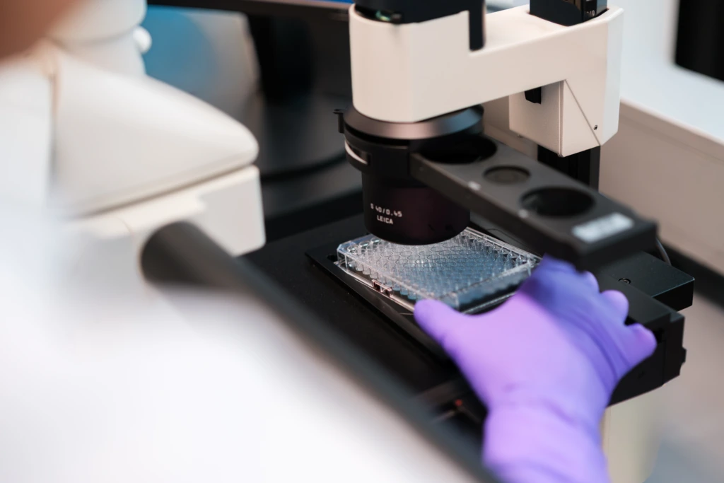
Our laboratories are set up to provide remote reading solutions to our customers. A fast, secure, and validated solution is used for the transferring of large image files. We offer remote viewing of images hosted on our own servers to avoid unnecessary downloading.
Having access to multiple software programs also provides extended capabilities to offer the most relevant and efficient method to generate data.
Our image analysis software capabilities includes:
We have designed a proprietary solution for automated adjudication and short turnaround time in clinical trials where regulatory authorities request multiple readers to analyze each biopsy.
Each pathologist is equipped with a dedicated tablet to scan the sample ID inserted into the biopsy image and report on the results directly. Scores are compared by an algorithm validated specifically for each trial/scenario, triggering the request for an additional reader when necessary. The single final consensus result is then reported in the clinical trial database.
This solution offers unequaled speed and reliability when multiple readings are required in a short time frame, like, for example, biopsies for the inclusion of patients in NASH trials.
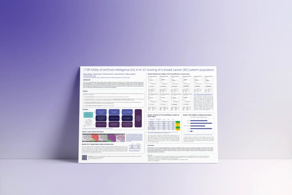
Discover how AI-powered image analysis enhances the accuracy and reproducibility of Ki-67 scoring for tumor proliferation assessment.
Explore how digital pathology and AI support standardized, quantitative assessment of proliferation markers in clinical oncology studies.
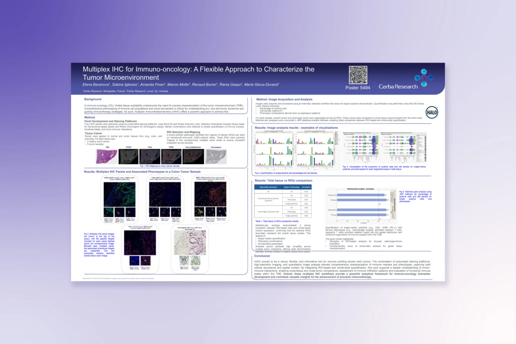
See how multiplex IHC and advanced image analysis enable detailed profiling of immune biomarkers in complex tumor environments.
Learn how digital pathology and image analysis support biomarker-driven approaches in non-small cell lung cancer.

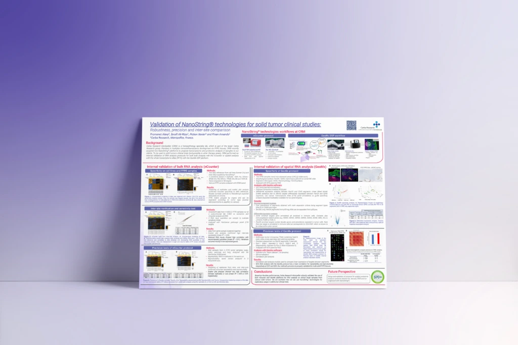
Explore how validated analytical approaches support robust, reproducible biomarker analysis in clinical tissue studies.
See how innovative digital pathology approaches improve quantification and interpretation of tissue changes in metabolic disease.