Get in touch with us
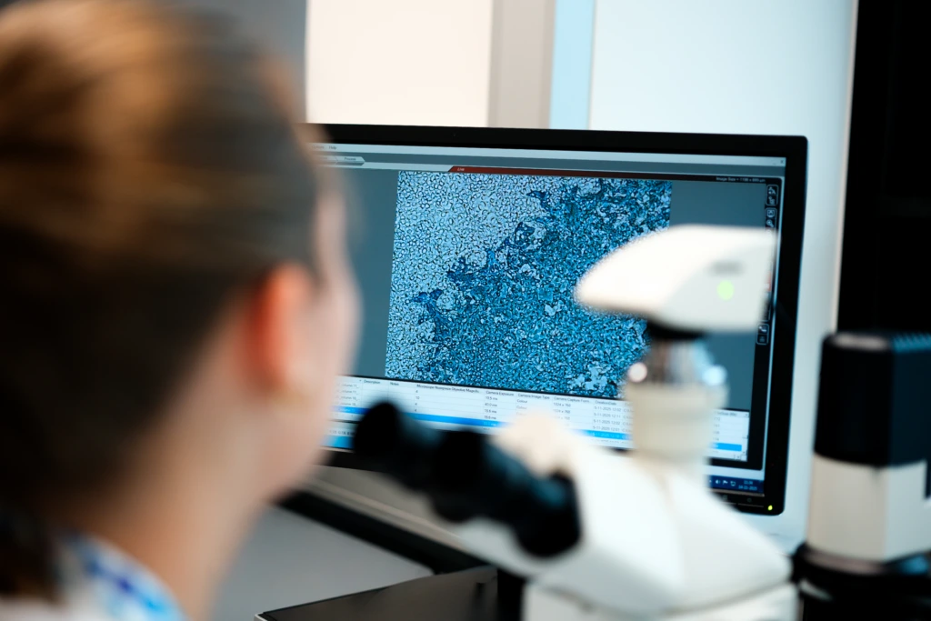
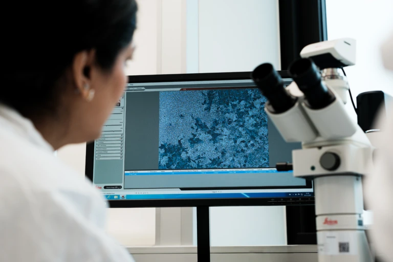
Scientist viewing microscope imaging results on monitor

Integrated Histopathology and Spatial Biology for Biomarker-Driven Clinical Development

Generate reproducible tissue biomarker data with integrated histopathology, digital pathology, and spatial biology solutions that support faster, more informed clinical development decisions.

Integrated expertise for complex tissue programs

Tissue analysis is central to translational and clinical research. driving deeper understanding of disease biology, treatment response, and clinically meaningful biomarkers throughout development.

Delivering high-quality histopathology data requires more than laboratory testing alone. It demands experienced pathologists, harmonized workflows, validated analytical approaches, and scientific teams who understand how tissue biomarkers align with study objectives, clinical endpoints, and regulatory expectations.

We combine histopathology expertise, immunohistochemistry (IHC), digital pathology, spatial biology, and advanced image analysis within a globally connected laboratory network built to support biomarker strategies across oncology, inflammation and immunology, neurology, metabolic disease, infectious disease, and rare disease programs.

From assay development and tissue qualification through to centralized pathology review and quantitative image analysis, we generate reproducible, clinically relevant tissue insights that help you reduce uncertainty, accelerate development, and move promising therapies closer to patients.

shutterstock_1634938366-1-scaled-1.jpg

Why choose Cerba Research for your histopathology programs?

Integrated tissue analysis expertise
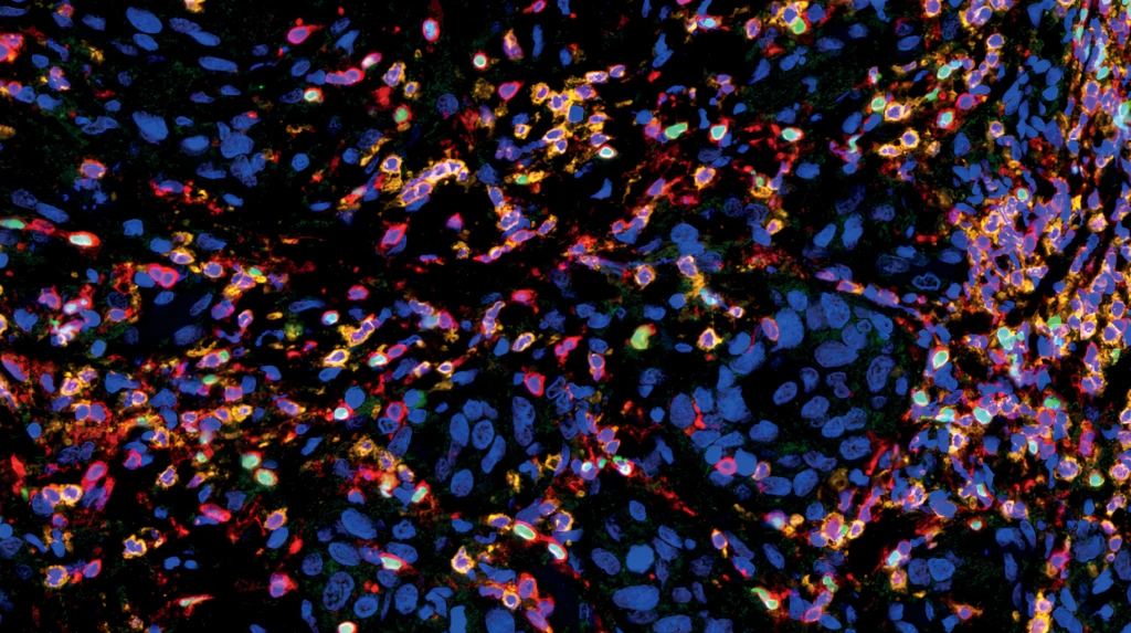
CD3 (Clone 2GV6, Red), CD8 (Clone AMC908, Orange) and FoxP3 (Clone 236A/E7, Green) multiplex IHC staining of Head and Neck Squamous Cell Carcinoma FFPE sample. T-Cells are identified by CD3 staining, Cytotoxic and regulatory subpopulations are identified by CD8 and FoxP3 staining respectively.
  • Histopathology, IHC, multiplex immunofluorescence, in situ hybridization (ISH), and digital pathology within one workflow
  • Scientific support from assay strategy through pathology interpretation
  • Experience supporting translational, registrational, and post-marketing studies

Data-driven insights at scale

Scientist reviewing microscope imaging data on monitor
  • Scalable digital pathology workflows enabling consistent tissue assessment 
  • Quantitative, reproducible biomarker analysis aligned to study endpoints
  • Expert-led integration of imaging, pathology, and data interpretation to support confident decision-making
  • Advanced imaging and analysis platforms supporting standardized and high-throughput tissue evaluation

Established biomarker capabilities

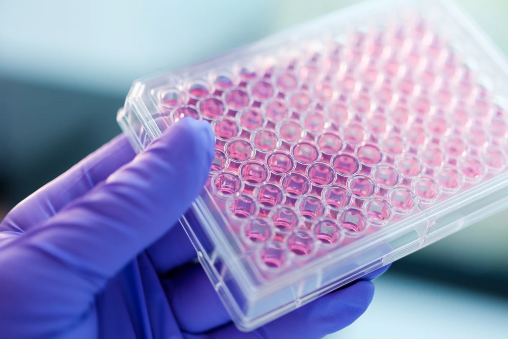
Gloved hand holding pink 96 well laboratory plate
  • ~160 validated IHC targets and 30+ multiplex panels available
  • Custom assay development, transfer, and fit-for-purpose validation support 
  • Multiplex and spatial biology workflows for complex biomarker strategies

Global pathology network

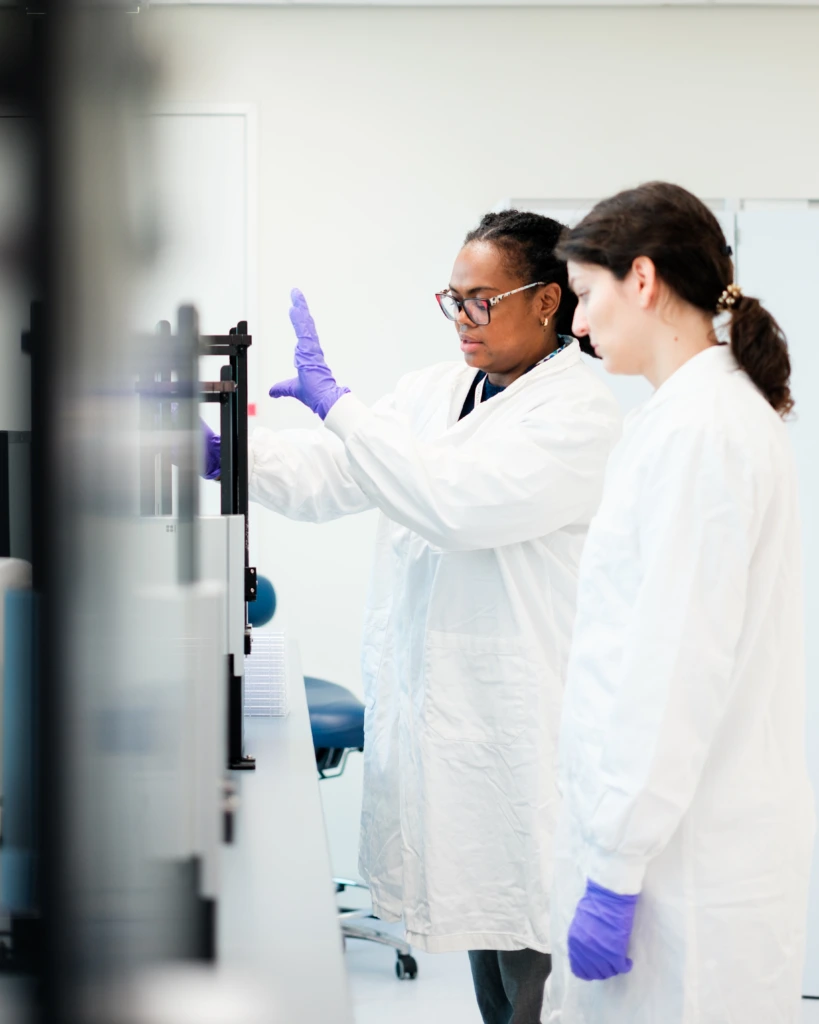
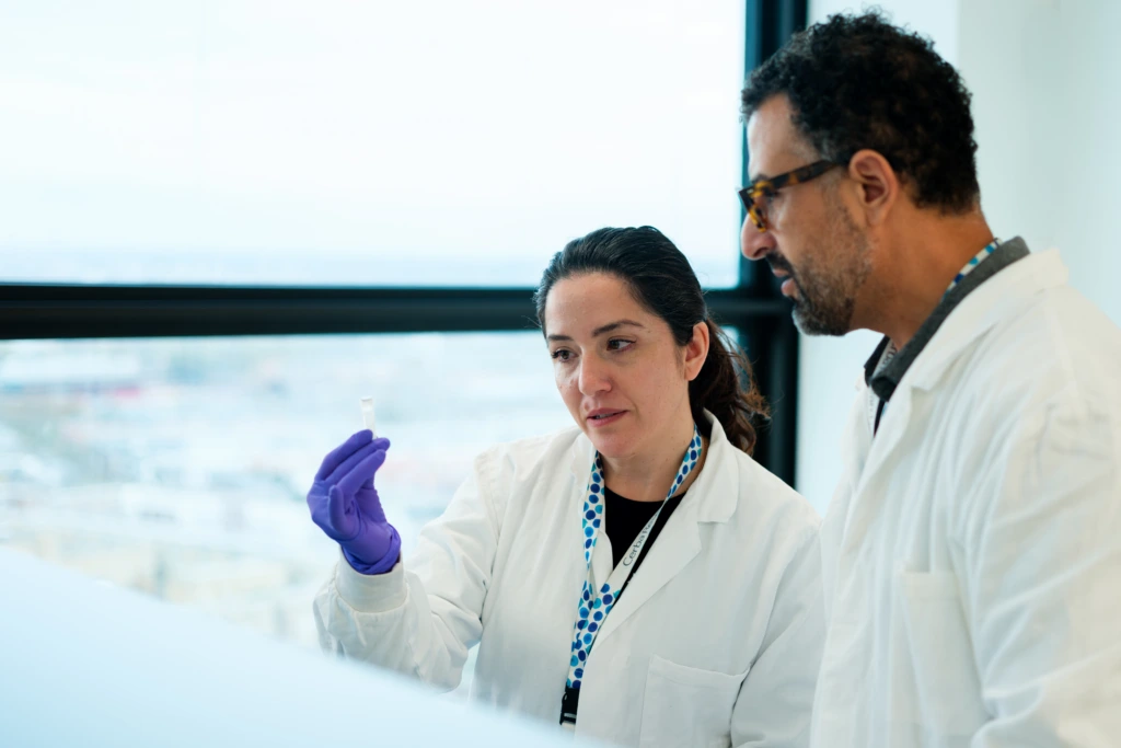
Scientists discussing clipboard beside laboratory workspace
  • Access to a network of 47 CerbaPath pathologists across 23 specialties
  • Additional external pathology expertise available for specialized study requirements
  • Harmonized processes and quality standards across regions
  • Centralized and remote pathology review capabilities

Supporting your histopathology programs

Our teams support your tissue biomarker strategy across every phase of clinical development, with integrated histopathology, molecular analysis, and digital workflows to deliver consistent, decision-ready data.

Immunohistochemistry and tissue biomarker analysis

Digital pathology and image analysis

Spatial biology and multiplex tissue analysis

Tissue sourcing and biorepository services

Immunohistochemistry and tissue biomarker analysis

Immunohistochemistry is a foundational tool for tissue biomarker characterization, supporting patient stratification, disease profiling, pharmacodynamic assessment, and translational research.

We work with you to develop, validate, and transfer tissue biomarker assays that align with your study objectives – from exploratory research through late-phase clinical trials. Whether supporting primary or secondary endpoints, standardized workflows and pathology-guided interpretation help ensure reproducible and clinically meaningful results across sites.

Our experience spans established and emerging biomarkers, including PD-L1, HER2, and ER/PR, supporting consistent application across oncology and other therapeutic areas.

Capabilities include:

  • Simplex and multiplex IHC
  • ~160 validated IHC targets and 30+ multiplex panels
  • Assay development and transfer
  • Sponsor-provided antibody qualification and optimization
  • Fit-for-purpose assay validation
  • Brightfield and fluorescence imaging
  • Automated staining platforms including Roche Ventana Discovery Ultra, Leica Biosystems Bond RX, and Agilent Dako Omnis
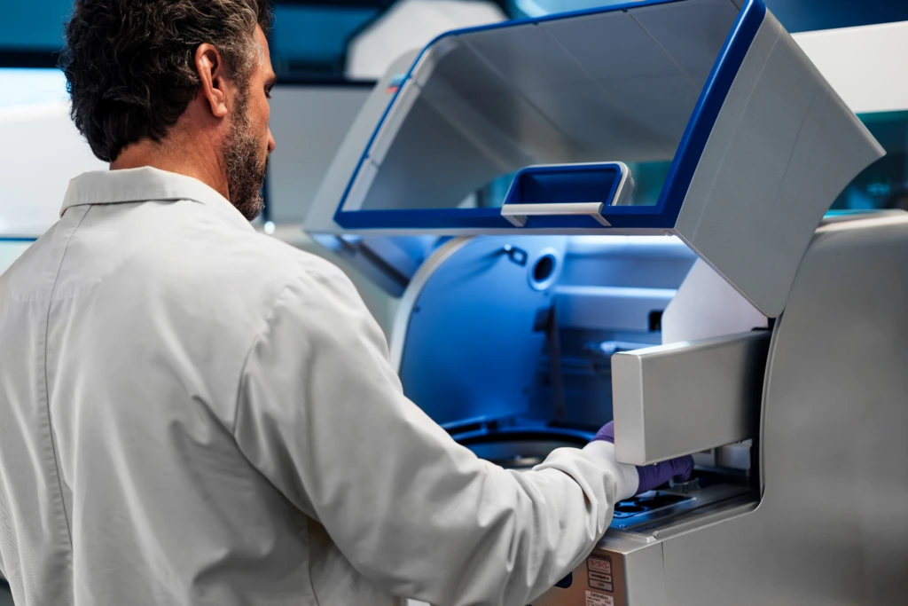
Scientists operating automated liquid handling system

Digital pathology and image analysis

Digital pathology enables consistent tissue assessment across geographically distributed clinical programs, helping to standardize interpretation and scale analysis across sites.

We combine pathology expertise with advanced imaging and analysis workflows to support centralized review, reduce variability, and deliver quantitative tissue data aligned with your study endpoints. This integrated approach helps ensure that complex tissue data is interpreted consistently and translated into decision-ready insights.

By aligning imaging, analysis, and pathology interpretation, we help you generate reproducible results that meet both scientific and regulatory expectations.

Capabilities include:

  • Whole-slide imaging and digital pathology workflows 
  • Centralized pathology review 
  • AI-assisted image analysis and tissue quantification 
  • Intra- and inter-observer variability assessments 
  • Hamamatsu and Akoya imaging platforms 
  • HALO® and Visiopharm® analysis platforms 
  • Multiparametric tissue analysis
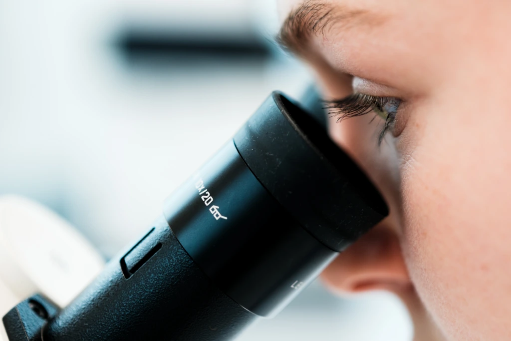
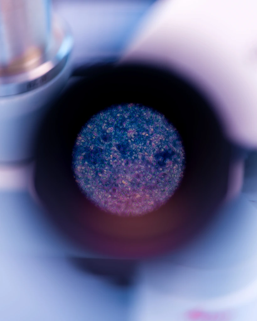
Close up of scientist using microscope eyepiece

Spatial biology and multiplex tissue analysis

Spatial biology approaches help characterize biomarker expression within the tissue microenvironment, providing additional context beyond single-marker analysis alone.

We support multiplex and spatial workflows that combine tissue morphology with protein and transcriptomic data, enabling a more complete view of tissue biology. This added context is critical when exploring mechanisms of action, resistance, and complex disease pathways that extend beyond single-marker analysis.

By integrating spatial and molecular data, you can generate deeper insight into tissue architecture while maintaining alignment with your study objectives.

Capabilities include:

  • Spatial transcriptomics and proteomics 
  • Multiplex immunofluorescence 
  • RNAscope® and ISH workflows 
  • Fluorescence in situ hybridization (FISH)
  • NanoString GeoMx® Digital Spatial Profiler applications 
  • Region-of-interest analysis with pathology support 
  • Integrated bioinformatics support
Scientists discussing work together in laboratory workspace

Tissue sourcing and biorepository services

Access to high-quality tissue samples is critical for biomarker development and translational research. Pathologist-reviewed sourcing and standardized sample management processes help ensure that samples are fit for purpose and aligned with your study requirements.

We support both retrospective and prospective studies with access to well-characterized tissue collections across multiple disease areas. Through rigorous qualification and quality control, we help you reduce variability at the sample level and generate more reliable downstream data.

This ensures that the tissue data generated across your program is both robust and clinically relevant.

Capabilities include:

  • Formalin-fixed paraffin-embedded (FFPE) and frozen tissue samples 
  • Human and animal tissue collections 
  • Tumor and matched normal tissue sourcing 
  • Tissue microarray construction 
  • Pathologist qualification and QC review 
  • Custom sourcing support
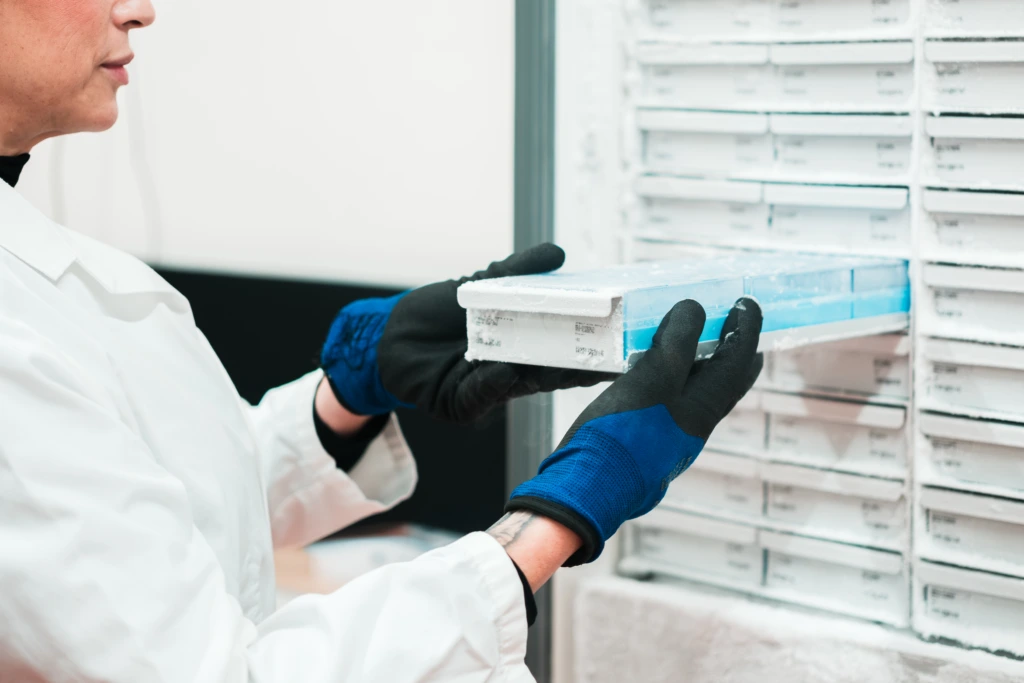
Female scientist accessing cold storage system B

Histopathology capabilities at a glance

~160
validated IHC targets
Duotone_Tick
30+
multiplex tissue analysis panels available
Duotone_Microscope
47
CerbaPath pathologists across 23 specialties
Duotone_Team-of-People

Global histopathology laboratories and pathology expertise

Work with globally connected histopathology laboratories that support consistent tissue analysis across your clinical programs, enabled by harmonized workflows and shared quality standards.

  • France (Frépillon)
  • China (Shanghai)
Frame_29_2x-2

Technologies used in histopathology programs

We use established histopathology, digital pathology, and spatial biology technologies to support reproducible tissue analysis across clinical development programs.

  • Automated IHC and staining platforms (Roche Ventana Discovery Ultra, Leica Biosystems Bond RX, Leica Biosystems Bond III, Agilent Dako Omnis, Roche Ventana Benchmark Ultra)
  • Special staining and histochemistry systems (Roche Ventana Benchmark Special Stains, Roche Ventana special stains, Agilent Dako Artisan Link Pro, Sakura Prisma Autostainer, Special Staining System)
  • Whole slide imaging and digital pathology scanners (Hamamatsu NanoZoomer® S360, Motic EasyScan Pro)
  • Digital pathology and image analysis software (Indica Labs HALO®, Visiopharm®, Akoya Biosciences inForm®)
  • Multiplex imaging and spatial biology platforms (Akoya Biosciences PhenoImager®, NanoString GeoMx® Digital Spatial Profiler)
  • In situ hybridization and RNA analysis (Advanced Cell Diagnostics RNAscope®)
  • Next-generation sequencing (NGS) platforms (Illumina NextSeq platform)
  • Gene expression and digital molecular profiling systems (NanoString nCounter® Analysis System)
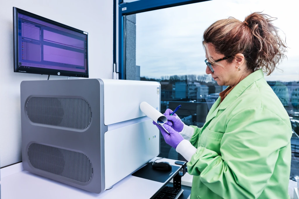
Medium shot of female scientist in green lab coat at laboratory analyser

Related specialized services

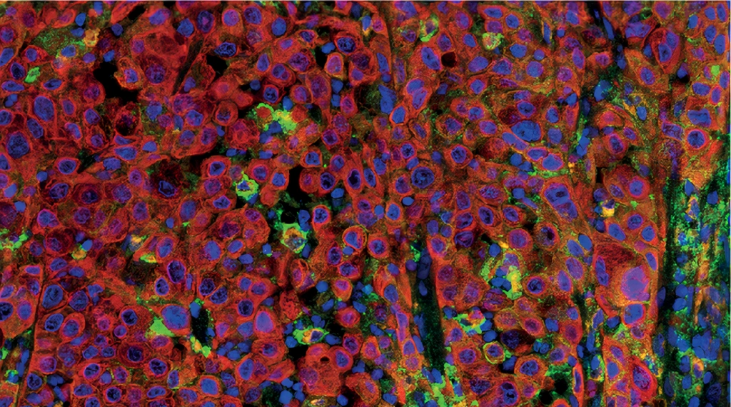
PanCK (Clone AE1/AE3, Red), CD68 (Clone KP1, Orange) and PD-L1 (Clone E1L3N, Green) multiplex IHC staining of Non-Small Cell Lung Cancer FFPE sample

Anatomic pathology and tissue analysis

Generate high-quality tissue data through integrated histopathology expertise, combining morphology, biomarker analysis, and pathology-led interpretation across clinical development.

Scientists collaborating at microscopy workstation

Digital imaging and analysis

Enable standardized and quantitative tissue assessment with digital pathology workflows, whole-slide imaging, and AI-assisted image analysis for reproducible results.

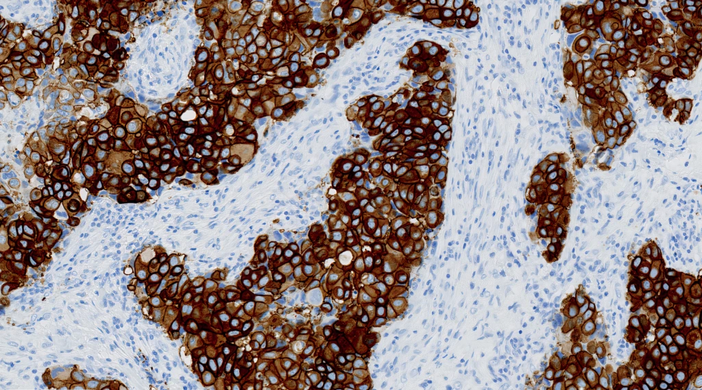
HercepTest™ mAB pharmDx Human Epidermal Growth Factor Receptor 2 (HER2) (Clone DG44) IHC staining of Breast Cancer  FFPE sample.

IHC and antibody services

Support biomarker characterization with robust immunohistochemistry solutions, including assay development, optimization, and validated antibody-based analysis.

Medium close up of scientist operating a laboratory analyzer machine A

Multiplex and hybridization imaging

Explore complex tissue biology using multiplex immunofluorescence and in situ hybridization techniques that enable multi-biomarker detection within tissue context.

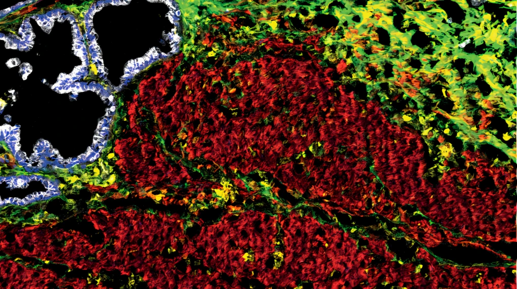
Representative image of 4 targets in a 12plex. Alpha-SMA (Clone 1A4, Red), Vimentin (Clone RV202, Yellow), PanCK (Clone C11, White), Collagen I (Polyclonal, Lime green) multiplex IMC staining of Frozen Pancreas sample. Platform: Discovery ULTRA.

Spatial omics

Characterize tissue biology with spatial transcriptomics and proteomics solutions that combine morphology with molecular insight.

Unlock the full value of your tissue data

From biomarker strategy to advanced pathology and image analysis, we help you generate reproducible insights that drive confident decisions across clinical development.

More histopathology insights

Poster

AI & Image Analysis for Ki-67 Quantification in Solid Tumors

Learn how AI-assisted image analysis improves reproducibility and quantitative accuracy of Ki-67 scoring in solid tumor tissue samples.

AI-and-Image-Analysis-for-Ki-67-Quantification-in-Solid-Tumors_Sun_Dark
Poster

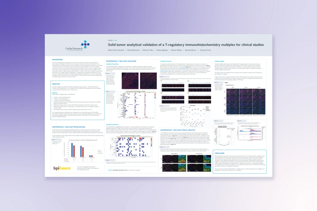
Analytical Validation of T-Regulatory IHC Multiplex in Solid Tumors

Understand how multiplex IHC assay validation supports robust, reproducible quantification of regulatory T-cell biomarkers in clinical trial tissue studies.

Cerba-Research-Poster-Solid-tumor-analytical-validation-of-a-T-regulatory-immunohistochemistry-multiplex-for-clinical-studies-1
Poster

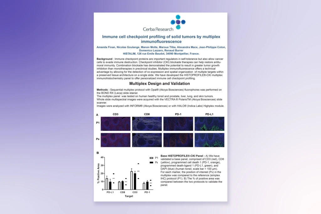
Immune Checkpoint Profiling of Solid Tumors via Multiplex IF

Explore how multiplex immunofluorescence enables detailed immune checkpoint profiling and deeper characterization of complex tumor immune microenvironments.

Immune Checkpoint Profiling of Solid Tumors via Multiplex Immunofluorescence_
Poster

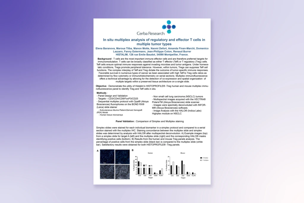
In Situ Multiplex Analysis of T Cells Across Multiple Tumor Types

Discover how spatially resolved multiplex analysis reveals regulatory and effector T-cell interactions within the tumor microenvironment across multiple tissue types.

In Situ Multiplex Analysis of T Cells Across Multiple Tumor Types
Whitepaper

The Value of Biomarkers from Discovery Through Commercialization

Learn how biomarkers support patient stratification, endpoint selection, and more informed decision-making across clinical development programs.

The Value of Biomarkers from Discovery Through Commercialization

Histopathology FAQ