
Get in touch with us
View all blogs

How to Validate Tumor Antigen Expression with Immunohistochemistry: Best Practices for Clinical Trials

Read the full blog

Before a therapy reaches patients, drug developers need to confirm that the antigen it targets is expressed in the right cells.

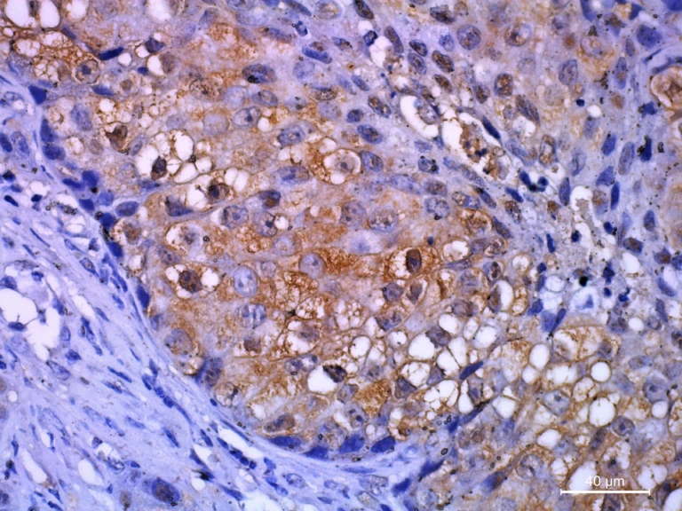
Immunohistochemistry (IHC), using highly specific monoclonal antibodies (mAbs), shows whether a tumor antigen is present, at what level, and whether the signal comes from malignant cells rather than surrounding stromal or inflammatory components.1,2 It’s why IHC remains a cornerstone of target validation in both diagnostic and therapeutic development.2

But the value of IHC depends on the assay. Variability in assay design, pre-analytical conditions, and interpretation can change which patients are selected for a trial and how clearly a therapy’s effect shows, particularly where tumors are heterogeneous within and between lesions.3

 Scoring systems and cutoffs also need to align with clinical endpoints and regulatory expectations, and in global trials, tissue handling, staining and interpretation must stay consistent from site to site. When they don’t, the impact reaches beyond assay variability to patient classification and the reading of therapeutic response, as HER2-low breast cancer and PD-L1 testing both show.5,6


Key challenges in IHC assay performance

HER2-low breast cancer: Misclassification due to assay sensitivity

In the past, IHC assays for HER2 grouped tumors into two categories: HER2-positive (IHC 3+ or amplified) and HER2-negative (IHC 0 or 1+). These assays were not designed to reliably distinguish between IHC 0 and 1+. Factors such as tissue fixation, antibody clone selection, and interpretation inconsistencies could lead to false negatives. 3 Some patients with true HER2 expression were mistakenly excluded from clinical trials. Furthermore, suboptimal IHC strategies diluted efficacy signals and underestimated treatment effects. Only after refining IHC strategies did HER2-low and indeed HER2-ultra low emerge as clinically actionable, for example with trastuzumab deruxtecan.4,5

PD-L1 expression: Assay variability and inconsistent patient selection

PD-L1 IHC, used as a companion diagnostic for immune checkpoint inhibitors, faces several challenges. Assays are not always analytically equivalent, and the same tumor sample can be defined as PD-L1 positive by one assay and negative by another.6 Heterogeneous and inducible PD-L1 expression further complicates interpretation. These issues can result in inappropriate exclusion of patients from therapy, weaker or inconsistent efficacy signals in trials, and unreliable cross-trial comparisons. Such challenges have been observed in non-small cell lung cancer (NSCLC) and triple-negative breast cancer (TNBC) trials. 6

In real-world scenarios, suboptimal IHC strategies can create erroneous biomarker profiles, ultimately obscuring true trial endpoints and weakening efficacy signals. This highlights the need for highly specific mAbs, rigorous assay validation, and standardized scoring systems.


Combatting common challenges with best practices in immunohistochemistry

To address these challenges, we promote best IHC practices to capture results that reflect true antigen expression, including intra-tumoral and inter-lesion heterogeneity. Key best practices include:

  1. Early alignment between discovery, translational, pathology, and clinical teams on target biology and assay purpose.
  2. Rigorous control of pre-analytical variables, including biopsy type, fixation time, and processing, with appropriate positive and negative controls.2
  3. Use of validated, well-characterized antibodies and scoring systems linked to clinically meaningful thresholds.
  4. Consideration of spatial and temporal heterogeneity through multiple sites and pre- and on-treatment biopsies to understand how expression evolves under therapy.
  5. Integration of IHC readouts with next-generation sequencing (NGS), flow cytometry, and PK/PD data to build a comprehensive view of target engagement and tumor microenvironment.


Good IHC practice supports:

  • Validation of antigen expression in relevant tumor subsets, such as HER2 and PD-L1.3
  • Assessment of spatial heterogeneity that can drive primary resistance or mixed responses.
  • Informing inclusion and exclusion criteria and biomarker-driven stratification in early and late-phase trials.
  • Complementing IHC with NGS and flow cytometry to provide spatial and microenvironment context to molecular and immune profiling data.


Key takeaways

  • IHC confirms target relevance: Precise antigen localization within tumor tissue supports confident validation of therapeutic targets and diagnostic markers.
  • Assay design directly impacts outcomes: Pre-analytical variables, antibody selection, and scoring approaches influence patient classification and trial data integrity.
  • Tumor heterogeneity complicates interpretation: Spatial and temporal variation in antigen expression can lead to misclassification and diluted efficacy signals.
  • Standardization improves consistency: Harmonized workflows across sites ensure reproducible staining, scoring, and cross-trial comparability.
  • Integrated biomarker strategies add depth: Combining IHC with NGS, flow cytometry, and PK/PD data enables a more complete understanding of tumor biology.
  • Optimized IHC enables better decisions: Robust validation and clinically aligned scoring support accurate patient selection and stronger trial outcomes.


Ready to strengthen your IHC strategy in oncology research?

When executed correctly, IHC confirms that a proposed antibody target is present in the right cells, at the right levels, and in the right spatial context before and during clinical development.1

If you’re developing targeted or immunotherapies, our integrated biomarker and assay development capabilities can help you generate reliable, clinically meaningful insights from every sample.

We combine expertise in IHC, biomarker testing, NGS, flow cytometry, and bioanalysis to help sponsors generate reliable data for target validation, patient selection, and treatment evaluation. Supporting modalities ranging from monoclonal antibodies and ADCs to bispecifics and cell therapies, our global laboratory network delivers the standardized workflows and scientific insight needed to take your program to the next phase.

Explore our oncology solutions or connect with our experts to see how we can support your next study.

References

  1. Köhler, G. and Milstein, C. 1975. Continuous cultures of fused cells secreting antibody of predefined specificity. Nature. 256(5517), pp.495-497.
  2. Shi, S.R., Cote, R.J. and Taylor, C.R. 1997. Antigen retrieval immunohistochemistry: past, present, and future. Journal of Histochemistry & Cytochemistry. 45(3), pp.327-343.
  3. Wolff, A.C., Hammond, M.E.H., Allison, K.H., Harvey, B.E., Mangu, P.B., Bartlett, J.M., Bilous, M., Ellis, I.O., Fitzgibbons, P., Hanna, W. and Jenkins, R.B. 2018. Human epidermal growth factor receptor 2 testing in breast cancer: American Society of Clinical Oncology/College of American Pathologists clinical practice guideline focused update. Archives of pathology & laboratory medicine. 142(11), pp.1364-1382.
  4. Modi, S., Gambhire, D. and Cameron, D. 2022. Trastuzumab deruxtecan in HER2-low breast cancer. Reply. The New England journal of medicine. 387(12), pp.1145-1146.
  5. Rakha, E.A., Tan, P.H., Van Bockstal, M.R., Allison, K.H., Brogi, E., Callagy, G., Cserni, G., Jaffer, S., Foschini, M.P., Gobbi, H. and Kulka, J. 2025. International expert consensus recommendations for HER2 reporting in breast cancer: focus on HER2-low and ultralow categories. Modern Pathology, p.100925.
  6. Hirsch, F.R., McElhinny, A., Stanforth, D., Ranger-Moore, J., Jansson, M., Kulangara, K., Richardson, W., Towne, P., Hanks, D., Vennapusa, B. and Mistry, A. 2017. PD-L1 immunohistochemistry assays for lung cancer: results from phase 1 of the blueprint PD-L1 IHC assay comparison project. Journal of Thoracic Oncology. 12(2), pp.208-222.

More oncology insights and resources

Blog

Read time – 7 mins November 5, 2025

Combining Flow Cytometry, IHC & NGS in Immuno-oncology: Essential Trio for Biomarker Success

Explore immuno-oncology biomarker strategies using flow cytometry, IHC, and NGS to improve tumor profiling, immune insights, and clinical trial outcomes.
Dna,Strand,And,Cancer,Cell,Oncology,Research,Concept,3d,Rendering
Guide

Read time – 14 mins January 25, 2025

A Best Practice Guide for Laboratory Services in Oncology Trials

Oncology trials are undergoing a transformative shift, developing therapies that directly target cancer cells and aim for more efficient treatments with a better safety profile. This approach aims to improve quality of life, extend progression-free survival rates for cancer patients, and increase overall survival.
Oncology-eBook_Amethyst
Blog

Read time – 8 mins March 11, 2026

How Custom Assays and Biobanking Drive Better Clinical Outcomes

One of the most significant challenges facing biopharmaceutical and biotechnology companies is translating complex science into meaningful clinical outcomes. Kirsty Maclean, Global Head of R&D, talks about how our integrated framework helps accelerate decision-making.

CD3 (Clone D7A6E, Red), CD16 (Clone EPR16784, Green), CD56  (Clone MRQ-42, Orange) multiplex IHC staining of Non-Small Cell Lung Cancer FFPE sample.