

The Life of a Flow Cytometry Sample
Christopher Rota, PhD
Scientist, Flow Cytometry
For our very first topic, let’s answer the most fundamental question that comes up in our conversations with new scientific partners: how does a flow cytometry assay actually work?
While FCM as a technology has grown rapidly, assays today still rely on the same fundamental experimental paradigm as they did sixty years ago. First, cells of interest are isolated from the sample, most commonly using centrifugation (which isolates cells from a mixture based on their density). Other techniques can also be applied to refine the sample further to just your cells of interest; a common example is performing red blood cell lysis, which enhances the detection of nucleated cells in whole blood samples.
Next, the isolated cells are resuspended in a suitable buffer solution and “stained” with a set of pre-defined labels (usually, fluorescently tagged antibodies). The stained cells are then washed to remove any excess labels. Depending on the kinds of biomarkers targeted, the cells may also need to be fixed (to immobilize and preserve the cellular components) and/or permeabilized () to allow labels to penetrate the cytoplasm and nucleus).
After staining and washing, the cells are loaded into the cytometer and “acquired” through the sample injection port (SIP for short). Within the cytometer, a high-pressure stream of fluid carries the stained cells to the interrogation point. Here, the cells are illuminated by an array of lasers of distinct wavelengths (colors) that excite the fluorescent labels on the cells, causing them to emit light of their own. The combined emitted light signals from the cells are captured by a set of detectors carefully tuned to record light within unique, specific sections of the visible light range. The signals from these detectors are then translated into discrete values corresponding to the intensity of the signal received. A full set of these values is recorded for each individual cell within the sample, which is why FCM data files can be quite large depending on how many labels and cells you have!
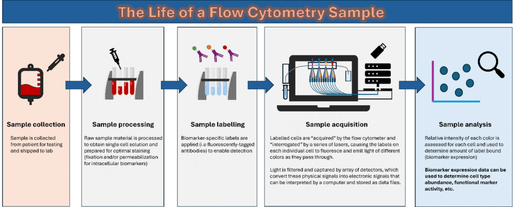
A visual overview of how flow cytometry assays are performed
This single cell recording is at the center of what makes flow cytometry so powerful. U; unlike other techniques that produce a single data point for a given biomarker for a group of cells (e.g an ELISA or traditional DNA/RNA sequencing), FCM can produce hundreds, thousands, or millions of data points from that same population. The sheer size of these data sets not only gives increased confidence in the data gathered for common cell types but also means that rare cells of interest can be identified with great precision (a common need in many clinical studies).
Using Flow Cytometry in Clinical Trial Research
Having gained a better understanding of the kind of data flow assays produce and how they produce it, a good next question would be: how and when should you use flow cytometry in a clinical trial?
While FCM can be applied to almost any clinical setting, it shines most in the following situations:
You care about the cells in your sample
This point may sound obvious, but it is important to remember! Flow cytometry requires a certain density of labels on a single particle to provide accurate detection; free-floating labels bound to soluble proteins cannot be “read” by the instrument as they are too dilute.
You care about the functional state of your cells
Most FCM-compatible labels bind to proteins, meaning flow cytometry is generally most effective at providing a snapshot of what cells are currently doing. If you care more about what your cells are gearing up to do, examining the genome and transcriptome using a DNA or RNA-sequencing approach will give a more accurate picture of that latent state. Another benefit of focusing on the proteome for FCM is that it can directly assess drug binding and persistence. This pharmacokinetic information is extremely useful in many applications but particularly valuable for immuno-oncology.
Your cells have a high degree of heterogeneity that is important to your study
Flow cytometry’s biggest strength is its ability to detect proteins with single cell resolution. For complex matrices like whole blood or bone marrow, being able to tease apart the complex milieu of cell types at work in the sample can yield essential insights about how each type of cell is changing over the course of a study. If you are looking for a cell type-specific response, flow cytometry is also one of the most sensitive techniques available for capturing that type of information. Many applications can benefit from this type of data, such as vaccine testing (which will also be discussed in a later post).

A quick quiz to help you decide if flow cytometry is right for your application
If one or more of the above are true for your study, then we would strongly urge you to consider flow cytometry as part of your portfolio of testing!
Join us next time for a deeper dive into one of the most prominent current applications for flow cytometry: studying cellular responses in immuno-oncology trials!

